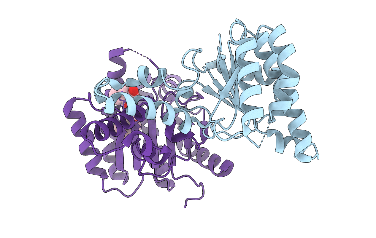
3SGT image
Entry Detail
PDB ID:
3SGT
Title:
Crystal Structure of E. coli undecaprenyl pyrophosphate synthase in complex with BPH-1299
Biological Source:
Source Organism:
Host Organism:
PDB Version:
Deposition Date:
2011-06-15
Release Date:
2012-12-19
Method Details:
Experimental Method:
Resolution:
1.85 Å
R-Value Free:
0.22
R-Value Work:
0.17
R-Value Observed:
0.17
Space Group:
P 21 21 21
Macromolecular Entities
Polymer Type:polypeptide(L)
Description:Undecaprenyl pyrophosphate synthase
Chain IDs:A (auth: B), B (auth: A)
Chain Length:253
Number of Molecules:2
Biological Source:Escherichia coli O6
Ligand Molecules
Primary Citation
Antibacterial drug leads targeting isoprenoid biosynthesis.
Proc.Natl.Acad.Sci.USA 110 123 128 (2013)
PMID: 23248302 DOI: 10.1073/pnas.1219899110

Abstact

With the rise in resistance to antibiotics such as methicillin, there is a need for new drugs. We report here the discovery and X-ray crystallographic structures of 10 chemically diverse compounds (benzoic, diketo, and phosphonic acids, as well as a bisamidine and a bisamine) that inhibit bacterial undecaprenyl diphosphate synthase, an essential enzyme involved in cell wall biosynthesis. The inhibitors bind to one or more of the four undecaprenyl diphosphate synthase inhibitor binding sites identified previously, with the most active leads binding to site 4, outside the catalytic center. The most potent leads are active against Staphylococcus aureus [minimal inhibitory concentration (MIC)(90) ∼0.25 µg/mL], and one potently synergizes with methicillin (fractional inhibitory concentration index = 0.25) and is protective in a mouse infection model. These results provide numerous leads for antibacterial development and open up the possibility of restoring sensitivity to drugs such as methicillin, using combination therapies.

Legend

Protein

Chemical

Disease

Primary Citation of related structures