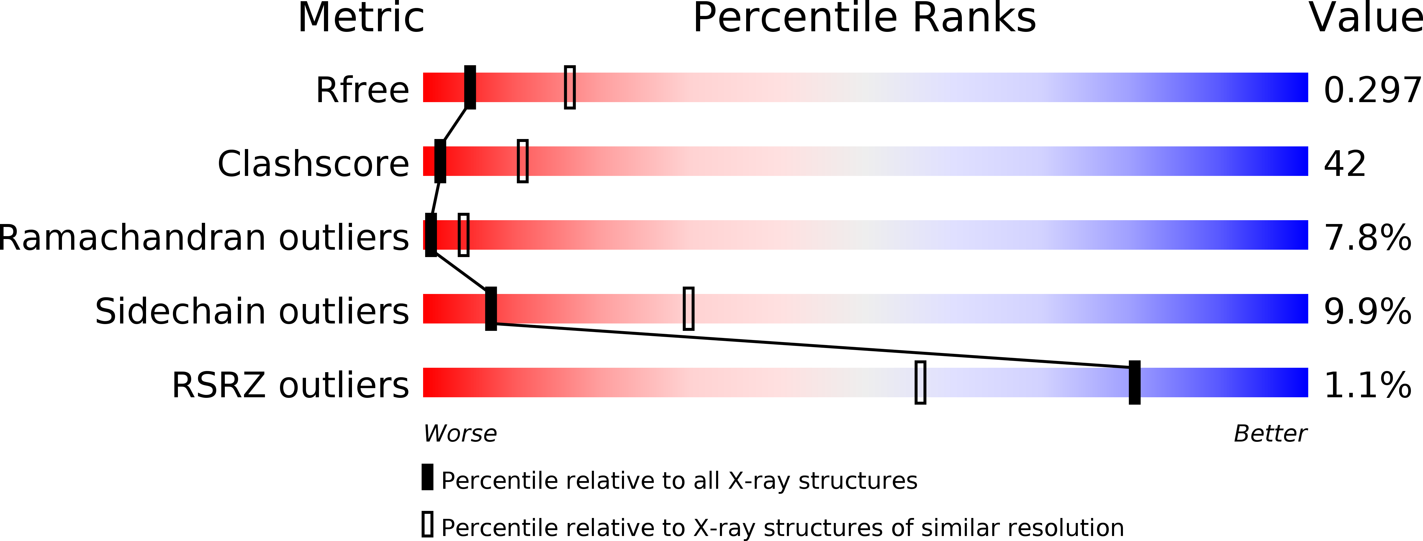
3SFZ image
Entry Detail
PDB ID:
3SFZ
Keywords:
Title:
Crystal structure of full-length murine Apaf-1
Biological Source:
Source Organism:
Host Organism:
PDB Version:
Deposition Date:
2011-06-14
Release Date:
2011-08-24
Method Details:
Experimental Method:
Resolution:
3.00 Å
R-Value Free:
0.29
R-Value Work:
0.23
R-Value Observed:
0.23
Space Group:
P 21 21 21
Macromolecular Entities
Polymer Type:polypeptide(L)
Description:Apoptotic peptidase activating factor 1
Chain IDs:A
Chain Length:1249
Number of Molecules:1
Biological Source:Mus musculus
Primary Citation
Crystal structure of full-length Apaf-1: how the death signal is relayed in the mitochondrial pathway of apoptosis.
Structure 19 1074 1083 (2011)
PMID: 21827944 DOI: 10.1016/j.str.2011.05.013

Abstact

The apoptotic protease-activating factor 1 (Apaf-1) relays the death signal in the mitochondrial pathway of apoptosis. Apaf-1 oligomerizes on binding of mitochondrially released cytochrome c into the heptameric apoptosome complex to ignite the downstream cascade of caspases. Here, we present the 3.0 Å crystal structure of full-length murine Apaf-1 in the absence of cytochrome c. The structure shows how the mammalian death switch is kept in its "off" position. By comparing the off state with a recent cryo-electron microscopy derived model of Apaf-1 in its apoptosomal conformation, we depict the molecular events that transform Apaf-1 from autoinhibited monomer to a building block of the caspase-activating apoptosome. Moreover, we have solved the crystal structure of the R265S mutant of full-length murine Apaf-1 in the absence of cytochrome c to 3.55 Å resolution and we show that proper function of Apaf-1 relies on R265 in the vicinity of the bound nucleotide.

Legend

Protein

Chemical

Disease

Primary Citation of related structures