3SFT image
Deposition Date 2011-06-14
Release Date 2011-07-20
Last Version Date 2023-11-01
Entry Detail
PDB ID:
3SFT
Keywords:
Title:
Crystal structure of Thermotoga maritima CheB methylesterase catalytic domain
Biological Source:
Source Organism:
Host Organism:
Method Details:
Experimental Method:
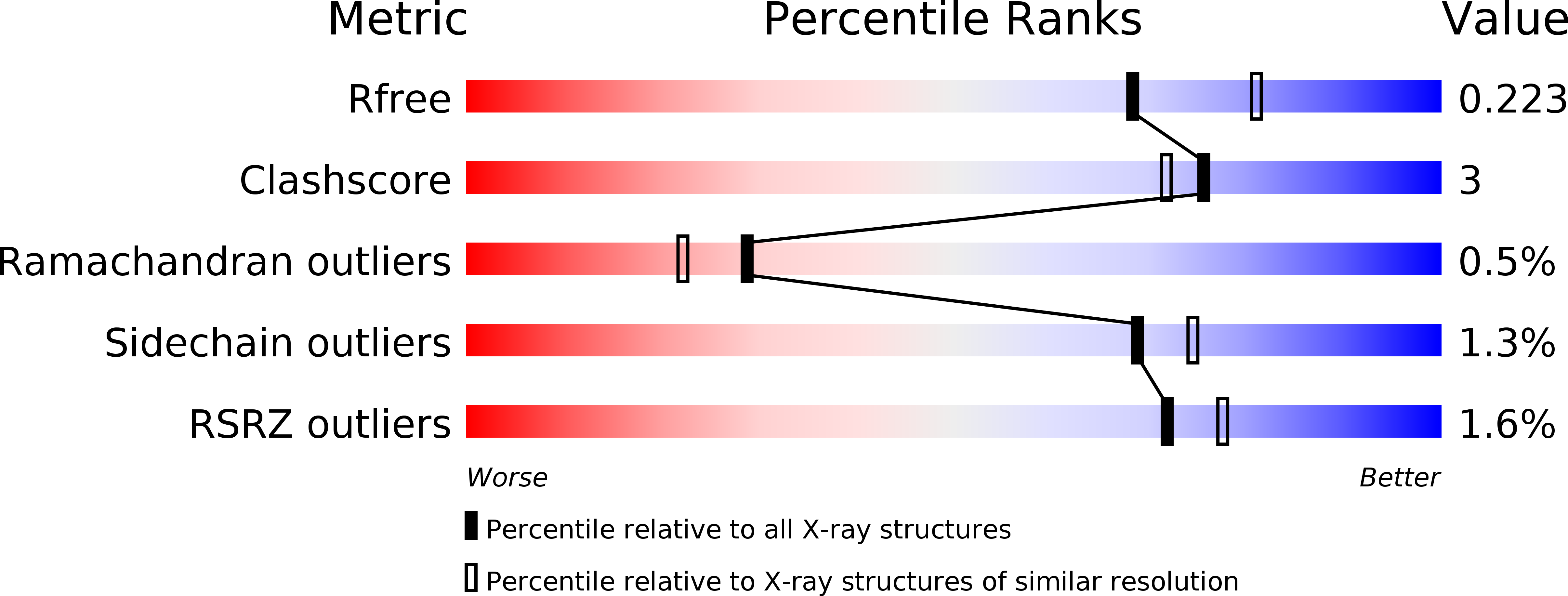
Resolution:
2.15 Å
R-Value Free:
0.22
R-Value Work:
0.20
Space Group:
P 21 21 21
Macromolecular Entities
Structures with similar UniProt ID
Protein Blast
Polymer Type:polypeptide(L)
Molecule:Chemotaxis response regulator protein-glutamate methylesterase
Gene (Uniprot):cheB
Chain IDs:A
Chain Length:193
Number of Molecules:1
Biological Source:Thermotoga maritima
Primary Citation
An insight into the interaction mode between CheB and chemoreceptor from two crystal structures of CheB methylesterase catalytic domain
Biochem.Biophys.Res.Commun. 411 69 75 (2011)
PMID: 21722627 DOI: 10.1016/j.bbrc.2011.06.090

Abstact

We have determined 2.2 Å resolution crystal structure of Thermotoga maritima CheB methylesterase domain to provide insight into the interaction mode between CheB and chemoreceptors. T. maritima CheB methylesterase domain has identical topology of a modified doubly-wound α/β fold that was observed from the previously reported Salmonella typhimurium counterpart, but the analysis of the electrostatic potential surface near the catalytic triad indicated considerable charge distribution difference. As the CheB demethylation consensus sites of the chemoreceptors, the CheB substrate, are not uniquely conserved between T. maritima and S. typhimurium, such surfaces with differing electrostatic properties may reflect CheB regions that mediate protein-protein interaction. Via the computational docking of the two T. maritima and S. typhimurium CheB structures to the respective T. maritima and Escherichia coli chemoreceptors, we propose a CheB:chemoreceptor interaction mode.

Legend

Protein

Chemical

Disease

Primary Citation of related structures
Feedback Form
Name
Email
Institute
Feedback