3SFM image
Deposition Date 2011-06-13
Release Date 2011-11-02
Last Version Date 2023-09-13
Entry Detail
PDB ID:
3SFM
Keywords:
Title:
Novel crystallization conditions for tandem variant R67 DHFR yields wild-type crystal structure
Biological Source:
Source Organism:
Escherichia coli (Taxon ID: 562)
Host Organism:
Method Details:
Experimental Method:
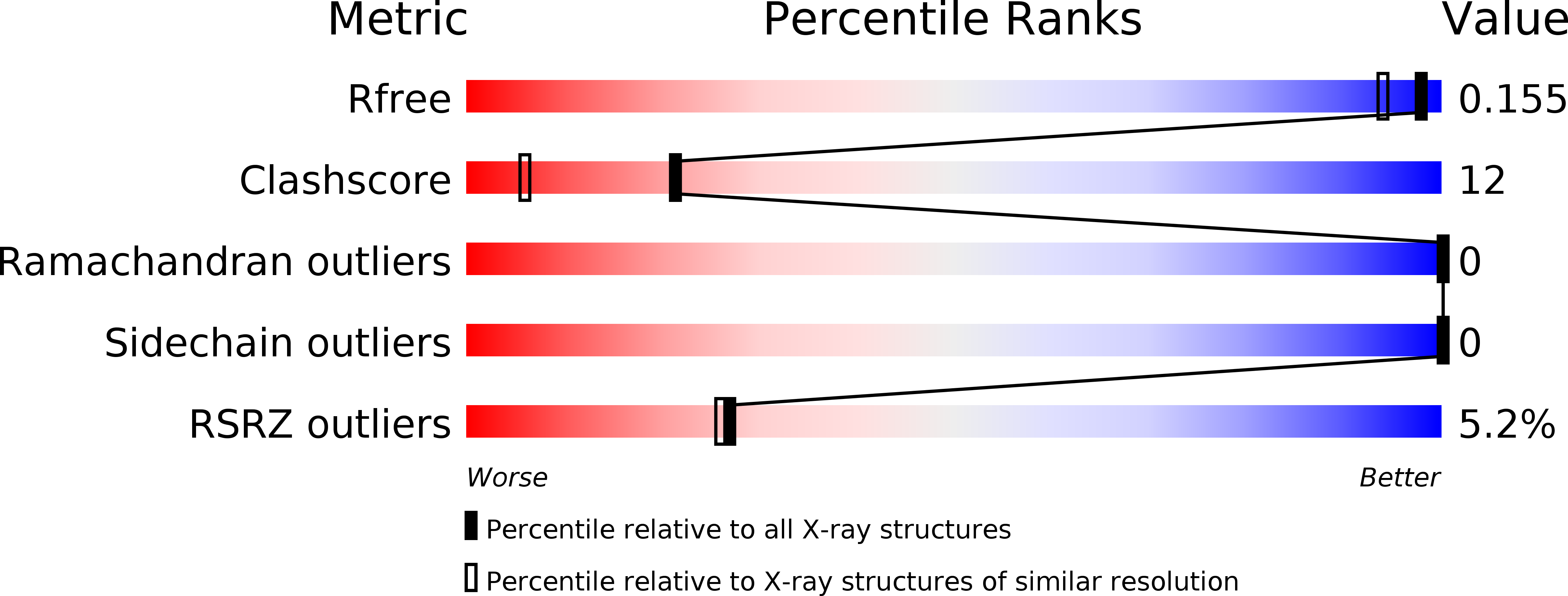
Resolution:
1.40 Å
R-Value Free:
0.16
R-Value Work:
0.14
R-Value Observed:
0.14
Space Group:
I 41 2 2
Macromolecular Entities
Polymer Type:polypeptide(L)
Molecule:Dihydrofolate reductase type 2
Chain IDs:A
Chain Length:62
Number of Molecules:1
Biological Source:Escherichia coli
Primary Citation
Novel crystallization conditions for tandem variant R67 DHFR yield a wild-type crystal structure.
Acta Crystallogr.,Sect.F 67 1316 1322 (2011)
PMID: 22102224 DOI: 10.1107/S1744309111030417

Abstact

Trimethoprim is an antibiotic that targets bacterial dihydrofolate reductase (DHFR). A plasmid-encoded DHFR known as R67 DHFR provides resistance to trimethoprim in bacteria. To better understand the mechanism of this homotetrameric enzyme, a tandem dimer construct was created that linked two monomeric R67 DHFR subunits together and mutated the sequence of residues 66-69 of the first subunit from VQIY to INSF. Using a modified crystallization protocol for this enzyme that included in situ proteolysis using chymotrypsin, the tandem dimer was crystallized and the structure was solved at 1.4 Å resolution. Surprisingly, only wild-type protomers were incorporated into the crystal. Further experiments demonstrated that the variant protomer was selectively degraded by chymotrypsin, although no canonical chymotrypsin cleavage site had been introduced by these mutations.

Legend

Protein

Chemical

Disease

Primary Citation of related structures