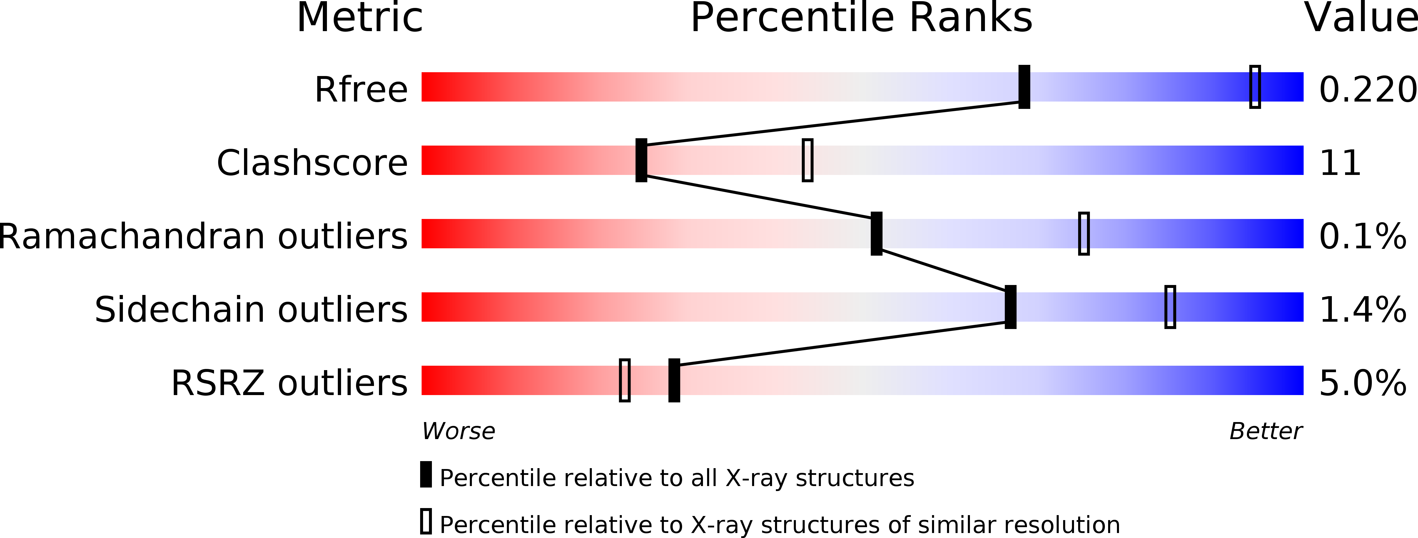
3SF4 image
Deposition Date 2011-06-12
Release Date 2011-11-23
Last Version Date 2023-11-01
Entry Detail
PDB ID:
3SF4
Title:
Crystal structure of the complex between the conserved cell polarity proteins Inscuteable and LGN
Biological Source:
Source Organism:
Homo sapiens (Taxon ID: 9606)
Host Organism:
Method Details:
Experimental Method:
Resolution:
2.60 Å
R-Value Free:
0.26
R-Value Work:
0.21
R-Value Observed:
0.21
Space Group:
P 41 21 2
Macromolecular Entities
Structures with similar UniProt ID
Protein Blast
Polymer Type:polypeptide(L)
Molecule:G-protein-signaling modulator 2
Gene (Uniprot):GPSM2
Chain IDs:A, B, C
Chain Length:406
Number of Molecules:3
Biological Source:Homo sapiens
Structures with similar UniProt ID
Protein Blast
Polymer Type:polypeptide(L)
Molecule:Protein inscuteable homolog
Gene (Uniprot):INSC
Chain IDs:D, E, F
Chain Length:52
Number of Molecules:3
Biological Source:Homo sapiens
Primary Citation
Structural basis for interaction between the conserved cell polarity proteins Inscuteable and Leu-Gly-Asn repeat-enriched protein (LGN)
Proc.Natl.Acad.Sci.USA 108 19210 19215 (2011)
PMID: 22074847 DOI: 10.1073/pnas.1110951108

Abstact

Interaction between the mammalian cell polarity proteins mInsc (mammalian homologue of Inscuteable) and Leu-Gly-Asn repeat-enriched protein (LGN), as well as that between their respective Drosophila homologues Inscuteable and Partner of Inscuteable (Pins), plays crucial roles in mitotic spindle orientation, a process contributing to asymmetric cell division. Here, we report a crystal structure of the LGN-binding domain (LBD) of human mInsc complexed with the N-terminal tetratricopeptide repeat (TPR) motifs of human LGN at 2.6-Å resolution. In the complex, mInsc-LBD adopts an elongated structure with three binding modules--an α-helix, an extended region, and a β-sheet connected with a loop--that runs antiparallel to LGN along the concave surface of the superhelix formed by the TPRs. Structural analysis and structure-based mutagenesis define residues that are critical for mInsc-LGN association, and reveal that the activator of G-protein signaling 3 (AGS3)-binding protein Frmpd1 [4.1/ezrin/radixin/moesin (FERM) and PSD-95/Dlg/ZO-1 (PDZ) domain-containing protein 1] and its relative Frmpd4 interact with LGN via a region homologous to a part of mInsc-LBD, whereas nuclear mitotic apparatus protein (NuMA) and the C terminus of LGN recognize the TPR domain in a manner different from that by mInsc. mInsc binds to LGN with the highest affinity (K(D) ≈ 2.4 nM) and effectively replaces the Frmpd proteins, NuMA, and the LGN C terminus, suggesting the priority of mInsc in binding to LGN. We also demonstrate, using mutant proteins, that mInsc-LGN interaction is vital for stabilization of LGN and for intracellular localization of mInsc.

Legend

Protein

Chemical

Disease

Primary Citation of related structures