Deposition Date
2011-06-09
Release Date
2012-03-14
Last Version Date
2024-02-28
Entry Detail
PDB ID:
3SDE
Keywords:
Title:
Crystal structure of a paraspeckle-protein heterodimer, PSPC1/NONO
Biological Source:
Source Organism(s):
Homo sapiens (Taxon ID: 9606)
Expression System(s):
Method Details:
Experimental Method:
Resolution:
1.90 Å
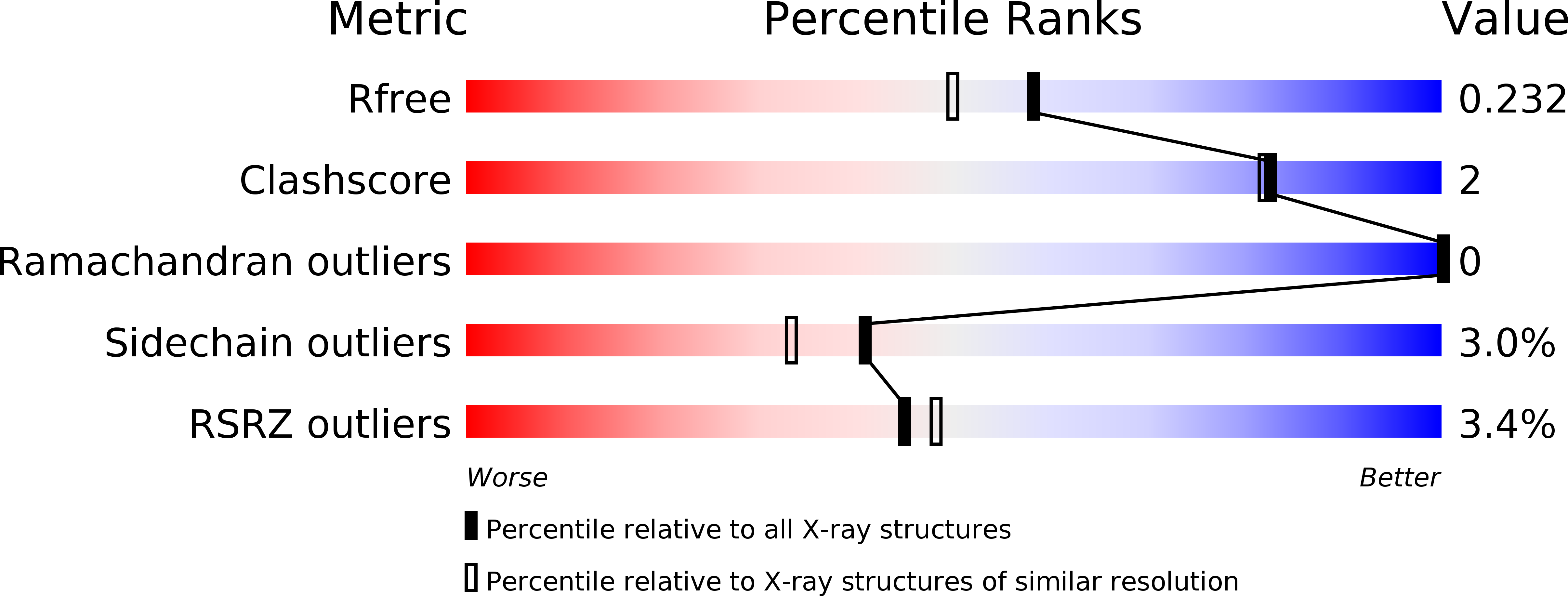
R-Value Free:
0.22
R-Value Work:
0.18
R-Value Observed:
0.18
Space Group:
C 1 2 1