3SC6 image
Deposition Date 2011-06-07
Release Date 2011-06-22
Last Version Date 2023-09-13
Entry Detail
PDB ID:
3SC6
Keywords:
Title:
2.65 Angstrom resolution crystal structure of dTDP-4-dehydrorhamnose reductase (rfbD) from Bacillus anthracis str. Ames in complex with NADP
Biological Source:
Source Organism:
Host Organism:
Method Details:
Experimental Method:
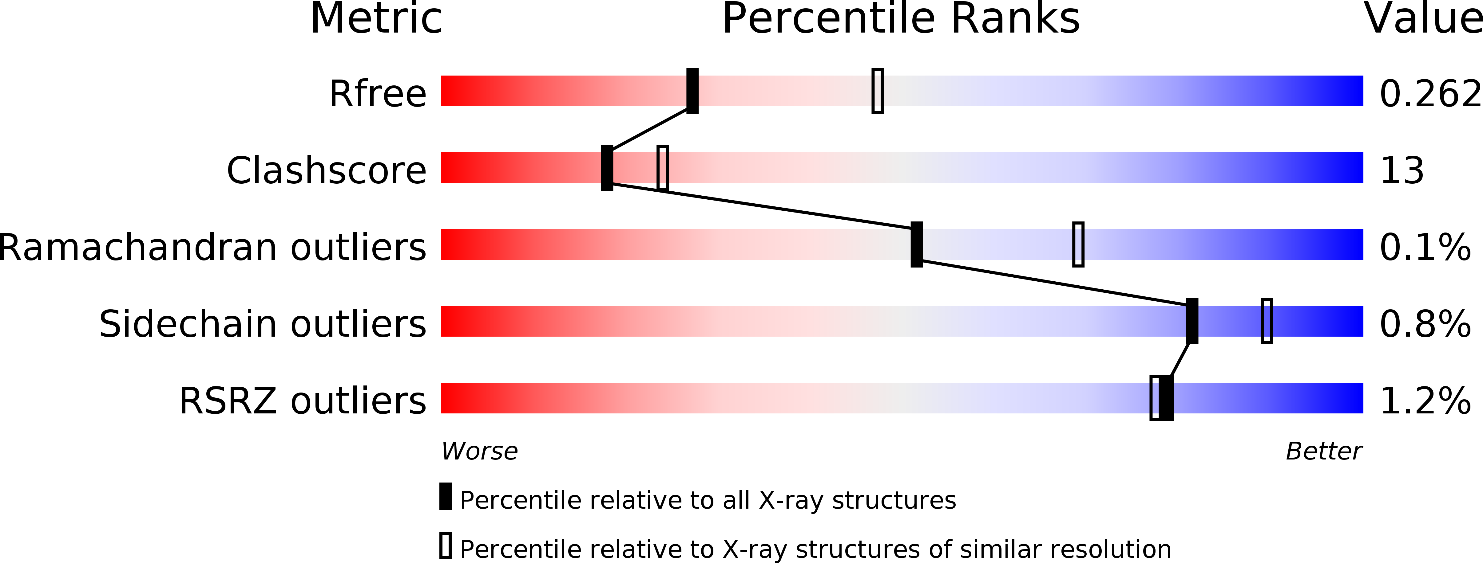
Resolution:
2.65 Å
R-Value Free:
0.25
R-Value Work:
0.21
R-Value Observed:
0.21
Space Group:
P 1 21 1
Macromolecular Entities
Structures with similar UniProt ID
Protein Blast
Polymer Type:polypeptide(L)
Molecule:dTDP-4-dehydrorhamnose reductase
Chain IDs:A, B, C, D, E, F
Chain Length:287
Number of Molecules:6
Biological Source:Bacillus anthracis
Primary Citation
Structure of the Bacillus anthracis dTDP-L-rhamnose-biosynthetic enzyme dTDP-4-dehydrorhamnose reductase (RfbD).
Acta Crystallogr F Struct Biol Commun 73 644 650 (2017)
PMID: 29199984 DOI: 10.1107/S2053230X17015746

Abstact

Bacillus anthracis is the causative agent of the deadly disease Anthrax. Its use in bioterrorism and its ability to re-emerge have brought renewed interest in this organism. B. anthracis is a Gram-positive bacterium that adds L-rhamnose to its cell-wall polysaccharides using the activated donor dTDP-β-L-rhamnose. The enzymes involved in the biosynthesis of the activated donor are absent in humans, which make them ideal targets for therapeutic development to combat pathogens. Here, the 2.65 Å resolution crystal structure of the fourth enzyme in the dTDP-β-L-rhamnose-biosynthetic pathway from B. anthracis, dTDP-4-dehydro-β-L-rhamnose reductase (RfbD), is presented in complex with NADP+. This enzyme catalyzes the reduction of dTDP-4-dehydro-β-L-rhamnose to dTDP-β-L-rhamnose. Although the protein was co-crystallized in the presence of Mg2+, the protein lacks the conserved residues that coordinate Mg2+.

Legend

Protein

Chemical

Disease

Primary Citation of related structures
Feedback Form
Name
Email
Institute
Feedback