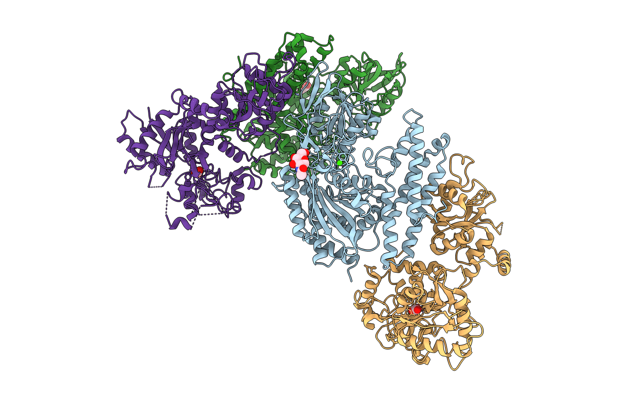
3S9L image
Entry Detail
PDB ID:
3S9L
Title:
Complex between transferrin receptor 1 and transferrin with iron in the N-Lobe, cryocooled 2
Biological Source:
Source Organism:
Host Organism:
PDB Version:
Deposition Date:
2011-06-01
Release Date:
2011-08-10
Method Details:
Experimental Method:
Resolution:
3.22 Å
R-Value Free:
0.31
R-Value Work:
0.27
Space Group:
P 43 2 2
Macromolecular Entities
Polymer Type:polypeptide(L)
Description:Transferrin receptor protein 1
Chain IDs:A, B
Chain Length:654
Number of Molecules:2
Biological Source:Homo sapiens
Polymer Type:polypeptide(L)
Description:Serotransferrin
Mutations:N427D,Y440F, Y531F N625D
Chain IDs:C, D
Chain Length:693
Number of Molecules:2
Biological Source:Homo sapiens
Modified Residue
Compound ID Chain ID Parent Comp ID Details 2D Image
ASN B ASN GLYCOSYLATION SITE
Primary Citation
How the binding of human transferrin primes the transferrin receptor potentiating iron release at endosomal pH.
Proc.Natl.Acad.Sci.USA 108 13089 13094 (2011)
PMID: 21788477 DOI: 10.1073/pnas.1105786108

Abstact

Delivery of iron to cells requires binding of two iron-containing human transferrin (hTF) molecules to the specific homodimeric transferrin receptor (TFR) on the cell surface. Through receptor-mediated endocytosis involving lower pH, salt, and an unidentified chelator, iron is rapidly released from hTF within the endosome. The crystal structure of a monoferric N-lobe hTF/TFR complex (3.22-Å resolution) features two binding motifs in the N lobe and one in the C lobe of hTF. Binding of Fe(N)hTF induces global and site-specific conformational changes within the TFR ectodomain. Specifically, movements at the TFR dimer interface appear to prime the TFR to undergo pH-induced movements that alter the hTF/TFR interaction. Iron release from each lobe then occurs by distinctly different mechanisms: Binding of His349 to the TFR (strengthened by protonation at low pH) controls iron release from the C lobe, whereas displacement of one N-lobe binding motif, in concert with the action of the dilysine trigger, elicits iron release from the N lobe. One binding motif in each lobe remains attached to the same α-helix in the TFR throughout the endocytic cycle. Collectively, the structure elucidates how the TFR accelerates iron release from the C lobe, slows it from the N lobe, and stabilizes binding of apohTF for return to the cell surface. Importantly, this structure provides new targets for mutagenesis studies to further understand and define this system.

Legend

Protein

Chemical

Disease

Primary Citation of related structures