Deposition Date
2011-06-01
Release Date
2011-09-07
Last Version Date
2024-10-09
Entry Detail
PDB ID:
3S9C
Keywords:
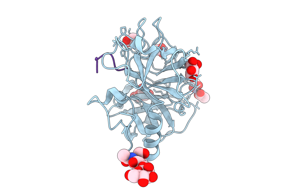
Title:
Russell's viper venom serine proteinase, RVV-V in complex with the fragment (residues 1533-1546) of human factor V
Biological Source:
Source Organism(s):
Homo sapiens (Taxon ID: 9606)
Daboia russellii siamensis (Taxon ID: 343250)
Daboia russellii siamensis (Taxon ID: 343250)
Method Details:
Experimental Method:
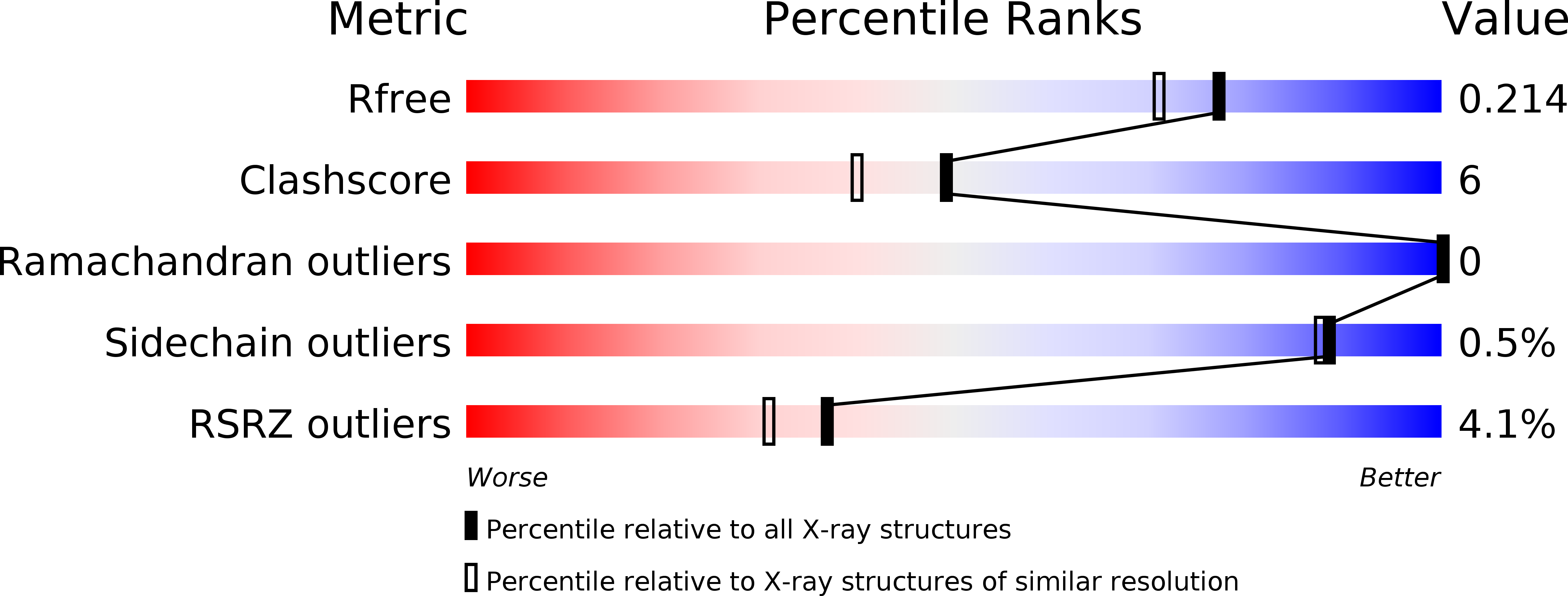
Resolution:
1.80 Å
R-Value Free:
0.21
R-Value Work:
0.18
R-Value Observed:
0.18
Space Group:
P 61