3S8Q image
Deposition Date 2011-05-30
Release Date 2012-01-18
Last Version Date 2024-02-28
Entry Detail
PDB ID:
3S8Q
Title:
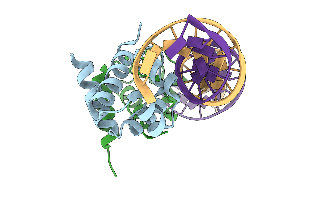
Crystal structure of the R-M controller protein C.Esp1396I OL operator complex
Biological Source:
Source Organism:
Host Organism:
Method Details:
Experimental Method:
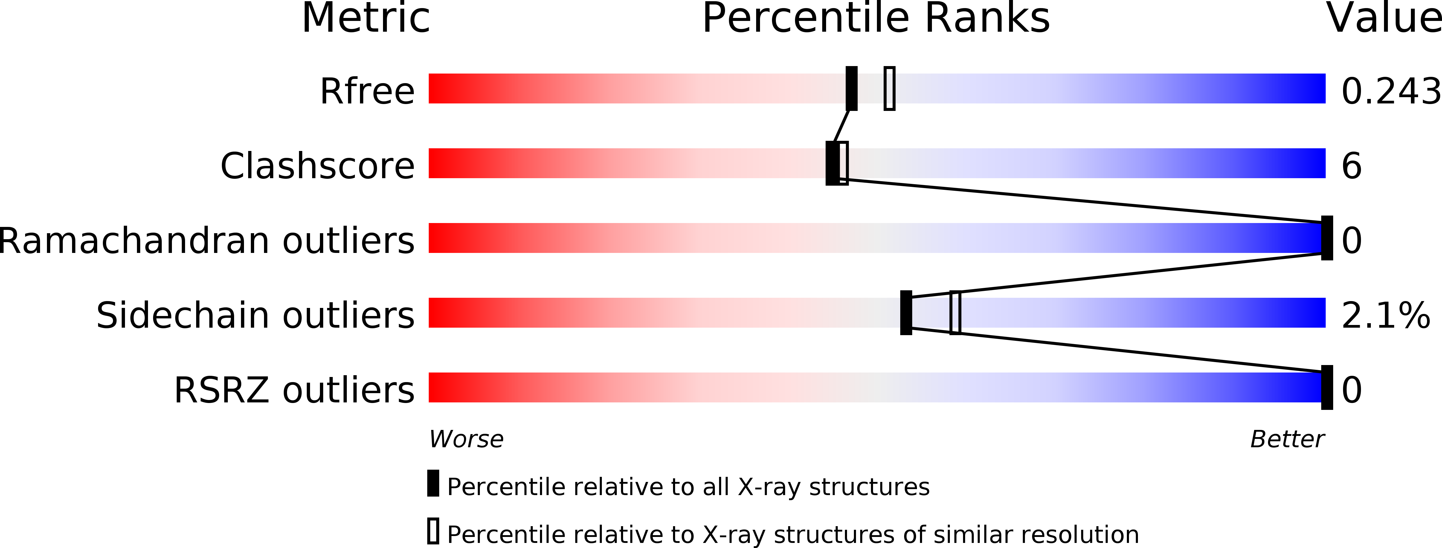
Resolution:
2.10 Å
R-Value Free:
0.21
R-Value Work:
0.15
R-Value Observed:
0.16
Space Group:
P 2 21 21
Macromolecular Entities
Structures with similar UniProt ID
Protein Blast
Polymer Type:polypeptide(L)
Molecule:R-M CONTROLLER PROTEIN
Gene (Uniprot):esp1396IC
Chain IDs:A, B
Chain Length:82
Number of Molecules:2
Biological Source:Enterobacter sp. RFL1396
Primary Citation
Recognition of dual symmetry by the controller protein C.Esp1396I based on the structure of the transcriptional activation complex.
Nucleic Acids Res. 40 4158 4167 (2012)
PMID: 22210861 DOI: 10.1093/nar/gkr1250

Abstact

The controller protein C.Esp1396I regulates the timing of gene expression of the restriction-modification (RM) genes of the RM system Esp1396I. The molecular recognition of promoter sequences by such transcriptional regulators is poorly understood, in part because the DNA sequence motifs do not conform to a well-defined symmetry. We report here the crystal structure of the controller protein bound to a DNA operator site. The structure reveals how two different symmetries within the operator are simultaneously recognized by the homo-dimeric protein, underpinned by a conformational change in one of the protein subunits. The recognition of two different DNA symmetries through movement of a flexible loop in one of the protein subunits may represent a general mechanism for the recognition of pseudo-symmetric DNA sequences.

Legend

Protein

Chemical

Disease

Primary Citation of related structures
Feedback Form
Name
Email
Institute
Feedback