Deposition Date
2011-05-26
Release Date
2011-10-05
Last Version Date
2023-09-13
Entry Detail
PDB ID:
3S7A
Keywords:
Title:
Human dihydrofolate reductase binary complex with PT684
Biological Source:
Source Organism(s):
Homo sapiens (Taxon ID: 9606)
Expression System(s):
Method Details:
Experimental Method:
Resolution:
1.80 Å
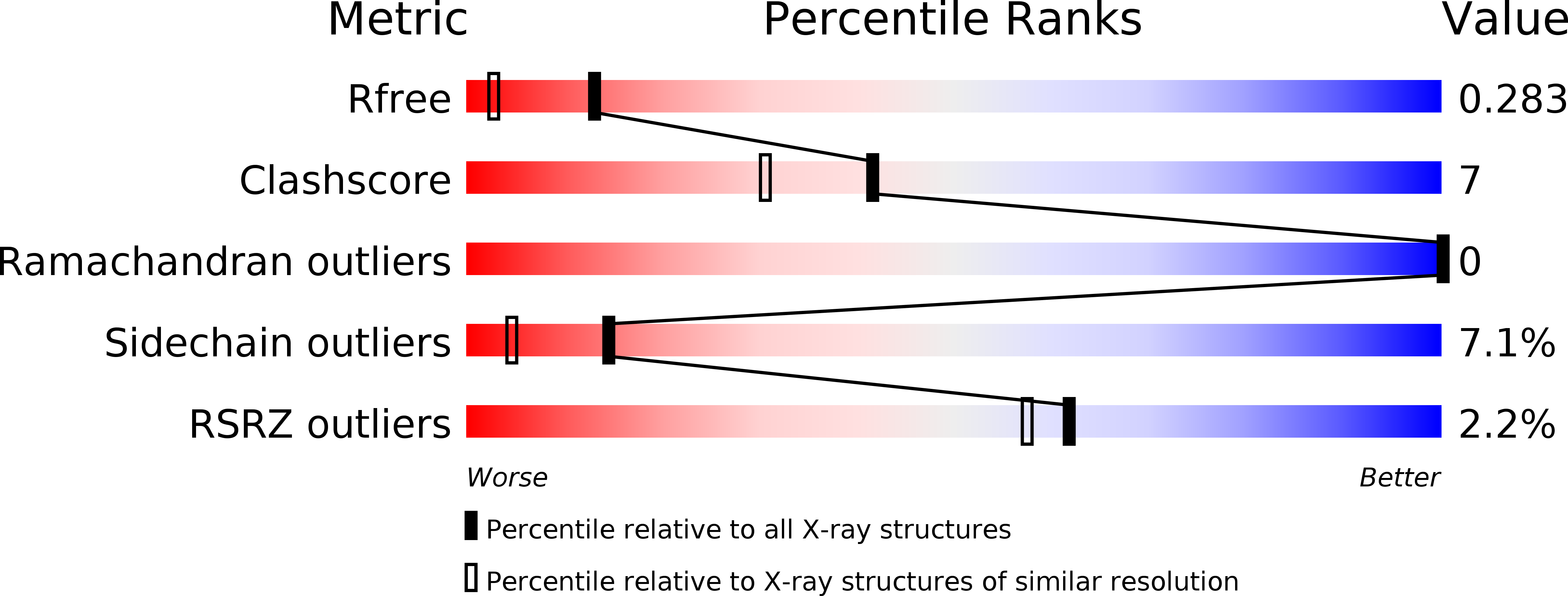
R-Value Free:
0.28
R-Value Work:
0.23
R-Value Observed:
0.23
Space Group:
P 21 21 21