3S5O image
Entry Detail
PDB ID:
3S5O
Keywords:
Title:
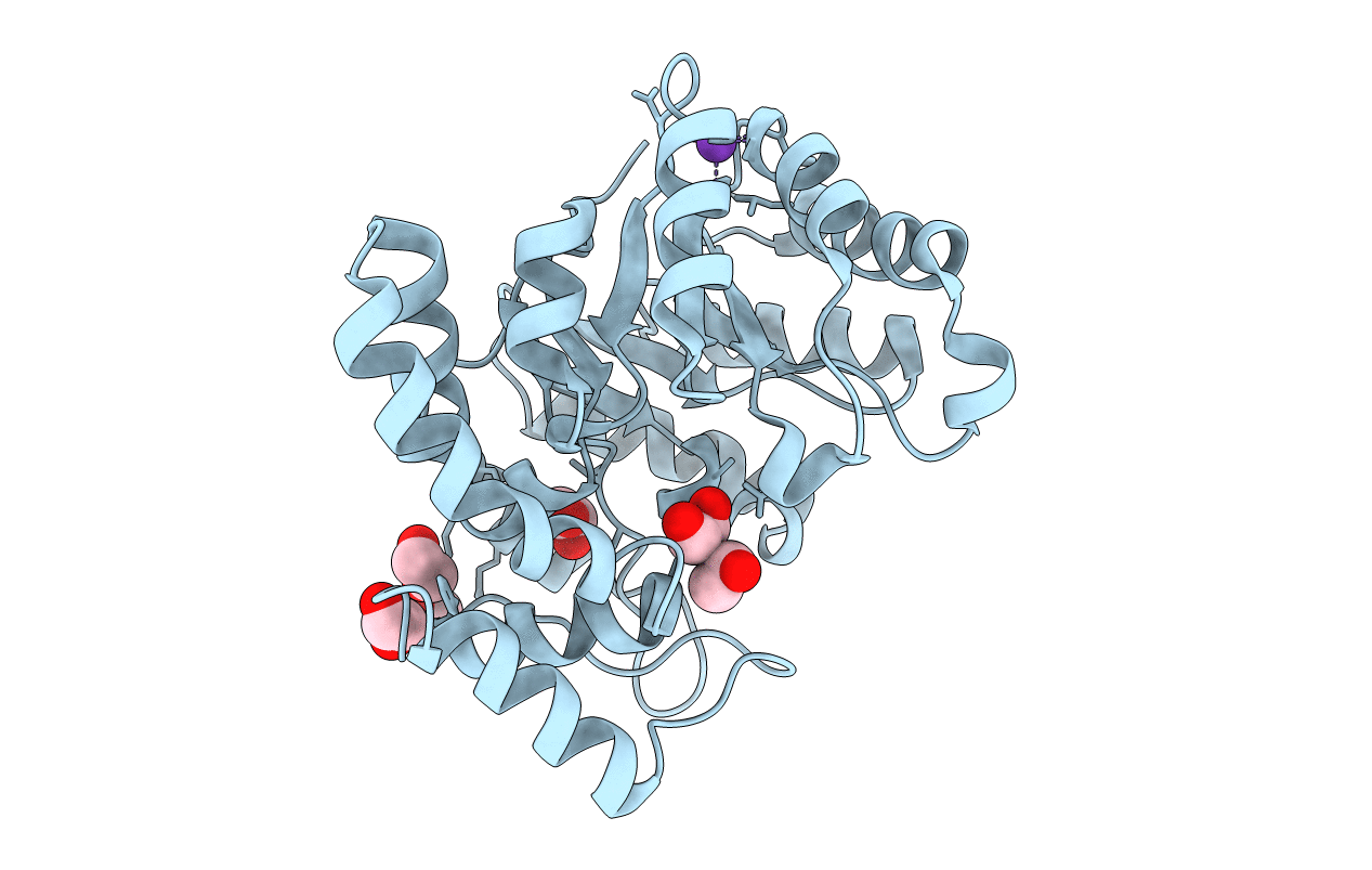
Crystal Structure of Human 4-hydroxy-2-oxoglutarate Aldolase Bound to Pyruvate
Biological Source:
Source Organism:
Host Organism:
PDB Version:
Deposition Date:
2011-05-23
Release Date:
2011-10-26
Method Details:
Experimental Method:
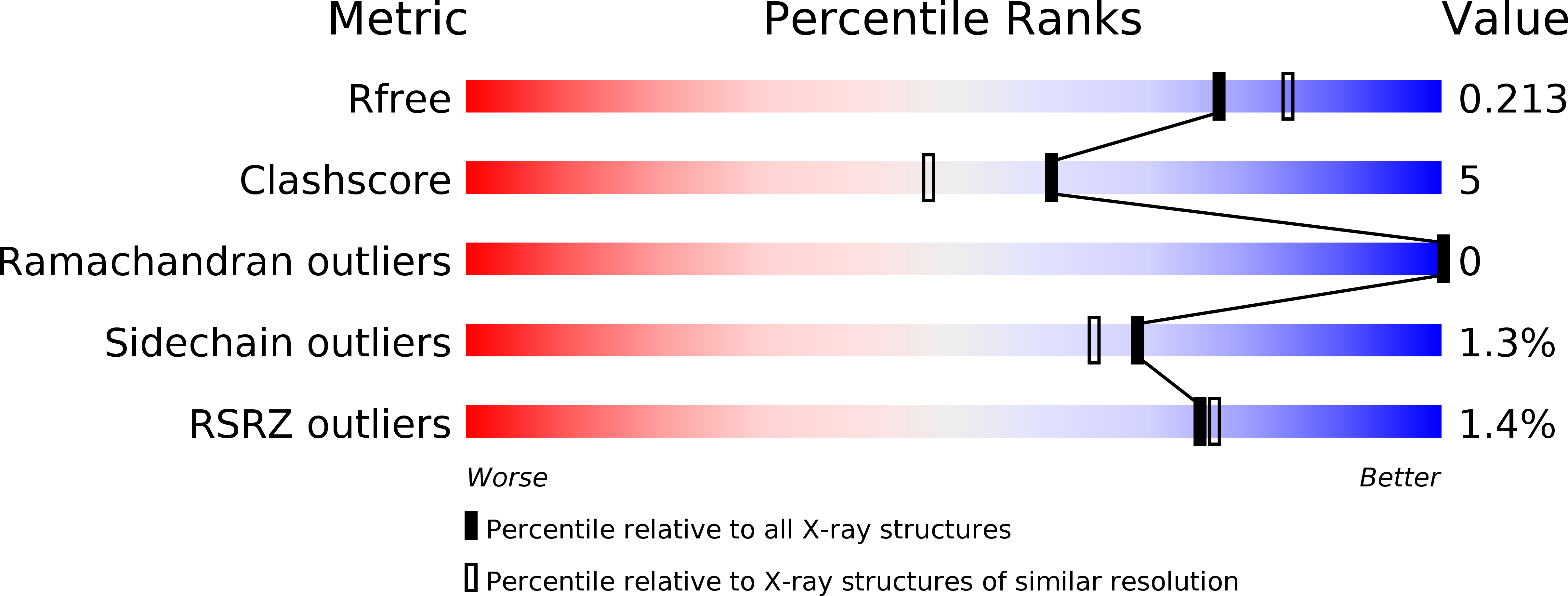
Resolution:
1.97 Å
R-Value Free:
0.21
R-Value Work:
0.19
R-Value Observed:
0.19
Space Group:
P 64 2 2
Macromolecular Entities
Polymer Type:polypeptide(L)
Description:4-hydroxy-2-oxoglutarate aldolase, mitochondrial
Chain IDs:A
Chain Length:307
Number of Molecules:1
Biological Source:Homo sapiens
Modified Residue
Compound ID Chain ID Parent Comp ID Details 2D Image
KPI A LYS ?
Primary Citation
Structural and Biochemical Studies of Human 4-hydroxy-2-oxoglutarate Aldolase: Implications for Hydroxyproline Metabolism in Primary Hyperoxaluria.
Plos One 6 e26021 e26021 (2011)
PMID: 21998747 DOI: 10.1371/journal.pone.0026021

Abstact

BACKGROUND 4-hydroxy-2-oxoglutarate (HOG) aldolase is a unique enzyme in the hydroxyproline degradation pathway catalyzing the cleavage of HOG to pyruvate and glyoxylate. Mutations in this enzyme are believed to be associated with the excessive production of oxalate in primary hyperoxaluria type 3 (PH3), although no experimental data is available to support this hypothesis. Moreover, the identity, oligomeric state, enzymatic activity, and crystal structure of human HOGA have not been experimentally determined. METHODOLOGY/PRINCIPAL FINDINGS In this study human HOGA (hHOGA) was identified by mass spectrometry of the mitochondrial enzyme purified from bovine kidney. hHOGA performs a retro-aldol cleavage reaction reminiscent of the trimeric 2-keto-3-deoxy-6-phosphogluconate aldolases. Sequence comparisons, however, show that HOGA is related to the tetrameric, bacterial dihydrodipicolinate synthases, but the reaction direction is reversed. The 1.97 Å resolution crystal structure of hHOGA bound to pyruvate was determined and enabled the modeling of the HOG-Schiff base intermediate and the identification of active site residues. Kinetic analyses of site-directed mutants support the importance of Lys196 as the nucleophile, Tyr168 and Ser77 as components of a proton relay, and Asn78 and Ser198 as unique residues that facilitate substrate binding. CONCLUSIONS/SIGNIFICANCE The biochemical and structural data presented support that hHOGA utilizes a type I aldolase reaction mechanism, but employs novel residue interactions for substrate binding. A mapping of the PH3 mutations identifies potential rearrangements in either the active site or the tetrameric assembly that would likely cause a loss in activity. Altogether, these data establish a foundation to assess mutant forms of hHOGA and how their activity could be pharmacologically restored.

Legend

Protein

Chemical

Disease

Primary Citation of related structures