3S5B image
Entry Detail
PDB ID:
3S5B
Keywords:
Title:
Crystal Structure of CED-3 Protease Suppressor-6 (CPS-6) from Caenorhabditis elegans
Biological Source:
Source Organism:
Host Organism:
PDB Version:
Deposition Date:
2011-05-23
Release Date:
2012-01-11
Method Details:
Experimental Method:
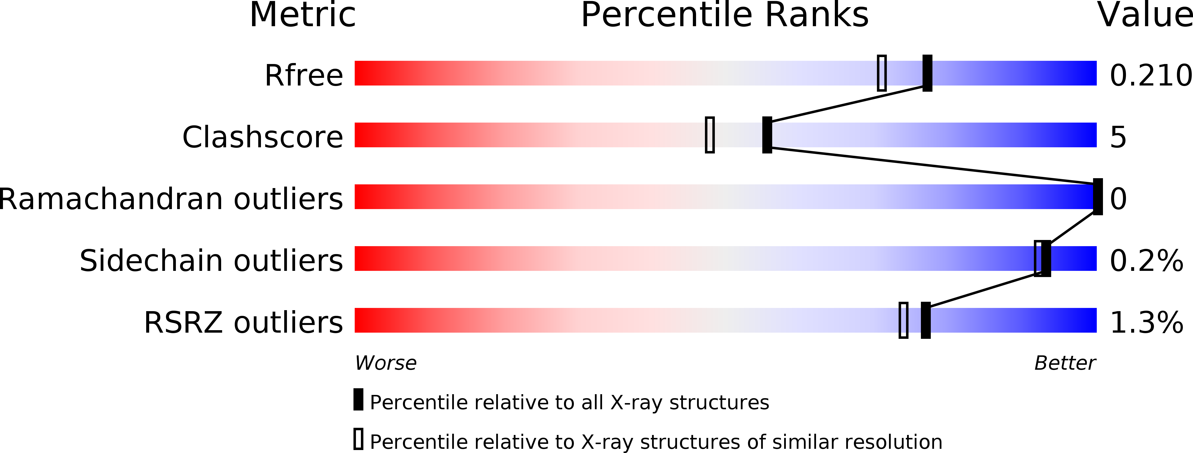
Resolution:
1.80 Å
R-Value Free:
0.20
R-Value Work:
0.16
R-Value Observed:
0.16
Space Group:
P 1 21 1
Macromolecular Entities
Polymer Type:polypeptide(L)
Description:Endonuclease G
Chain IDs:A, B
Chain Length:249
Number of Molecules:2
Biological Source:Caenorhabditis elegans
Ligand Molecules
Primary Citation
Structural insights into apoptotic DNA degradation by CED-3 protease suppressor-6 (CPS-6) from Caenorhabditis elegans
J.Biol.Chem. 287 7110 7120 (2012)
PMID: 22223640 DOI: 10.1074/jbc.M111.316075

Abstact

Endonuclease G (EndoG) is a mitochondrial protein that traverses to the nucleus and participates in chromosomal DNA degradation during apoptosis in yeast, worms, flies, and mammals. However, it remains unclear how EndoG binds and digests DNA. Here we show that the Caenorhabditis elegans CPS-6, a homolog of EndoG, is a homodimeric Mg(2+)-dependent nuclease, binding preferentially to G-tract DNA in the optimum low salt buffer at pH 7. The crystal structure of CPS-6 was determined at 1.8 Å resolution, revealing a mixed αβ topology with the two ββα-metal finger nuclease motifs located distantly at the two sides of the dimeric enzyme. A structural model of the CPS-6-DNA complex suggested a positively charged DNA-binding groove near the Mg(2+)-bound active site. Mutations of four aromatic and basic residues: Phe(122), Arg(146), Arg(156), and Phe(166), in the protein-DNA interface significantly reduced the DNA binding and cleavage activity of CPS-6, confirming that these residues are critical for CPS-6-DNA interactions. In vivo transformation rescue experiments further showed that the reduced DNase activity of CPS-6 mutants was positively correlated with its diminished cell killing activity in C. elegans. Taken together, these biochemical, structural, mutagenesis, and in vivo data reveal a molecular basis of how CPS-6 binds and hydrolyzes DNA to promote cell death.

Legend

Protein

Chemical

Disease

Primary Citation of related structures