3S4Z image
Deposition Date 2011-05-20
Release Date 2011-07-27
Last Version Date 2024-02-28
Entry Detail
PDB ID:
3S4Z
Title:
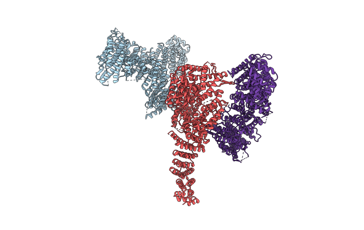
Structure of a Y DNA-FANCI complex
Biological Source:
Source Organism:
Mus musculus (Taxon ID: 10090)
Host Organism:
Method Details:
Experimental Method:
Resolution:
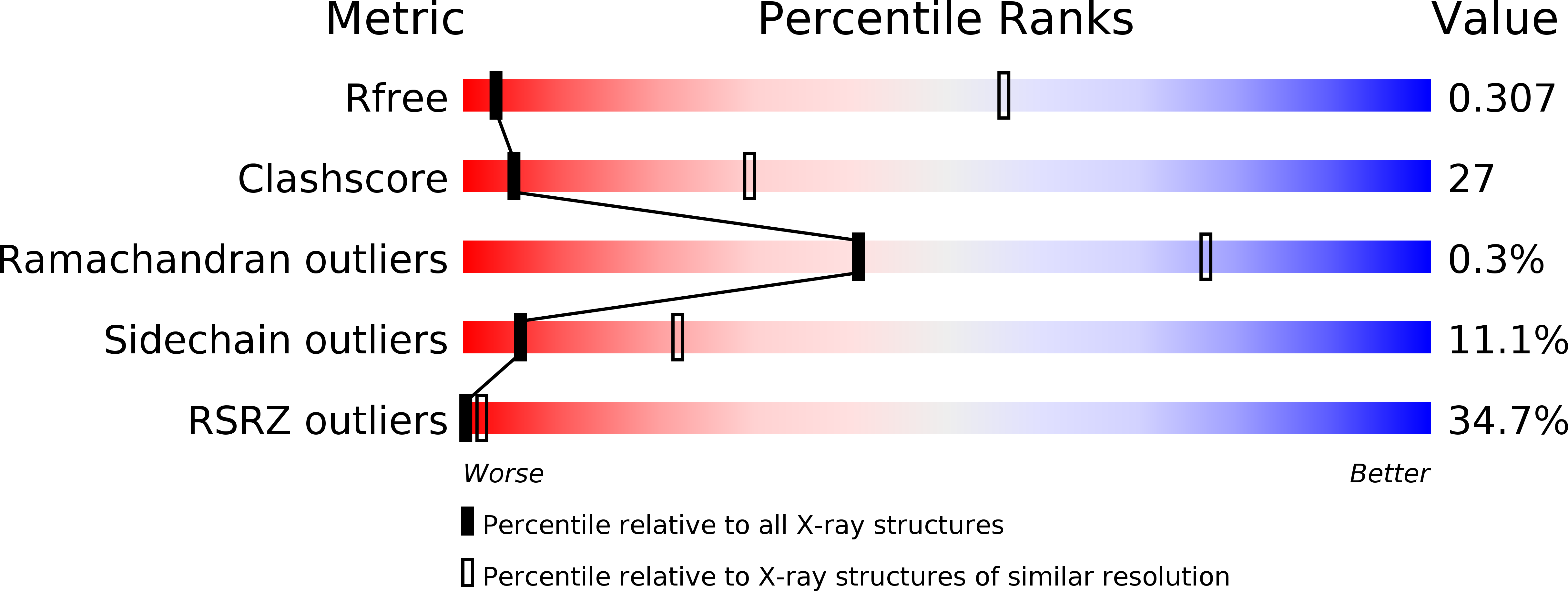
7.80 Å
R-Value Free:
0.32
R-Value Work:
0.30
R-Value Observed:
0.31
Space Group:
I 21 21 21
Macromolecular Entities
Polymer Type:polypeptide(L)
Molecule:dna repair 1
Gene (Uniprot):Fanci
Chain IDs:A, B, C
Chain Length:1308
Number of Molecules:3
Biological Source:Mus musculus
Ligand Molecules
Primary Citation
Structure of the FANCI-FANCD2 complex: insights into the Fanconi anemia DNA repair pathway.
Science 333 312 316 (2011)
PMID: 21764741 DOI: 10.1126/science.1205805

Abstact

Fanconi anemia is a cancer predisposition syndrome caused by defects in the repair of DNA interstrand cross-links (ICLs). Central to this pathway is the Fanconi anemia I-Fanconi anemia D2 (FANCI-FANCD2) (ID) complex, which is activated by DNA damage-induced phosphorylation and monoubiquitination. The 3.4 angstrom crystal structure of the ~300 kilodalton ID complex reveals that monoubiquitination and regulatory phosphorylation sites map to the I-D interface, suggesting that they occur on monomeric proteins or an opened-up complex and that they may serve to stabilize I-D heterodimerization. The 7.8 angstrom electron-density map of FANCI-DNA crystals and in vitro data show that each protein has binding sites for both single- and double-stranded DNA, suggesting that the ID complex recognizes DNA structures that result from the encounter of replication forks with an ICL.

Legend

Protein

Chemical

Disease

Primary Citation of related structures