3S4X image
Deposition Date 2011-05-20
Release Date 2012-08-01
Last Version Date 2023-09-13
Entry Detail
PDB ID:
3S4X
Keywords:
Title:
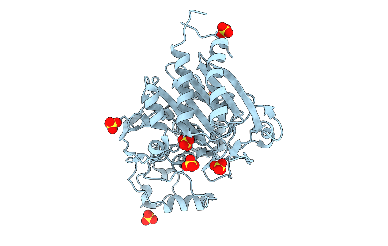
Crystal structure of the Asn152Gly mutant of P99 beta-lactamase
Biological Source:
Source Organism:
Host Organism:
Method Details:
Experimental Method:
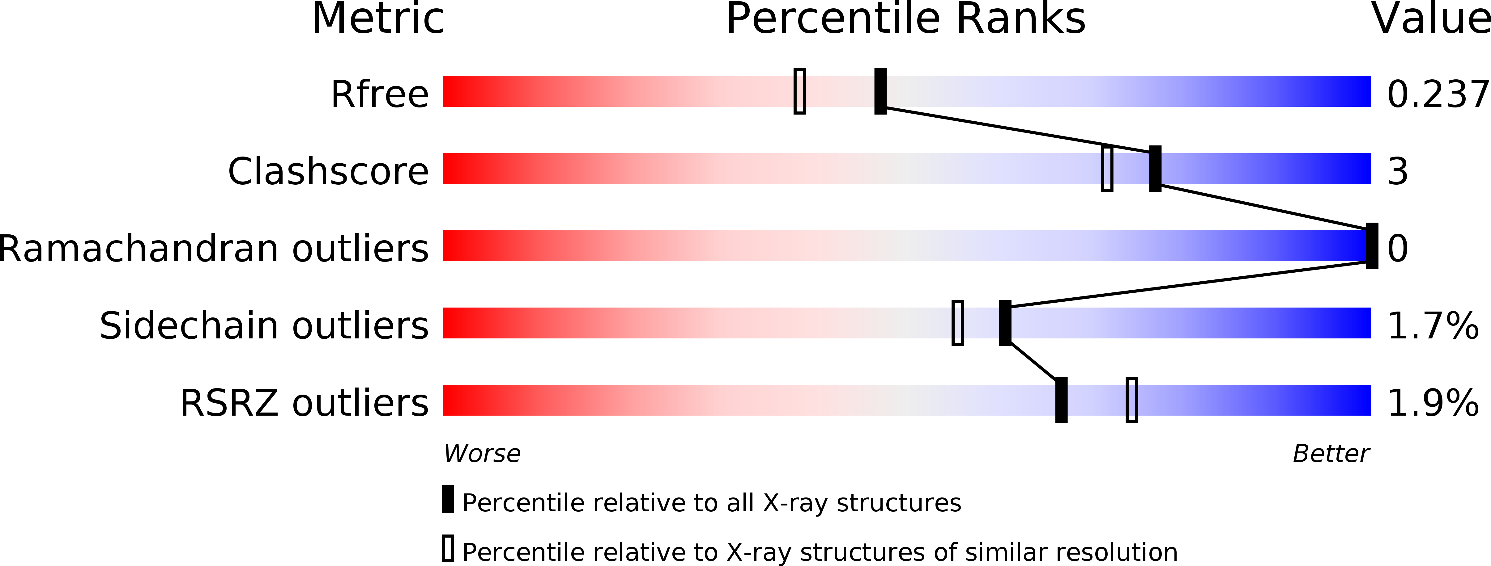
Resolution:
1.95 Å
R-Value Free:
0.23
R-Value Work:
0.17
R-Value Observed:
0.17
Space Group:
P 21 21 2
Macromolecular Entities
Polymer Type:polypeptide(L)
Molecule:Beta-lactamase
Gene (Uniprot):ampC
Mutations:I16V, A88P, N152G, A299V
Chain IDs:A
Chain Length:367
Number of Molecules:1
Biological Source:Enterobacter cloacae
Ligand Molecules
Primary Citation
Structural analysis of the Asn152Gly mutant of P99 cephalosporinase.
Acta Crystallogr.,Sect.D 68 1189 1193 (2012)
PMID: 22948919 DOI: 10.1107/S0907444912024080

Abstact

P99 cephalosporinase is a class C β-lactamase that is responsible in part for the widespread bacterial resistance to β-lactam antibiotics. Mutations of the conserved active-site residue Asn152 of the enzyme have been shown to alter β-lactam substrate specificity in vivo. Mutation of Asn152 to a glycine is notable in that it exhibits in vivo substrate-selectivity switching. In order to better understand the structural basis for this observed switch, the X-ray crystal structure of the apo Asn152Gly mutant of P99 was determined to 1.95 Å resolution. Unexpectedly, the artificial C-terminal His(6) tag of a symmetrically-related molecule was observed bound in the active site. The His(6) tag makes several interactions with key active-site residues, as well as with several sulfate ions. Additionally, the overall C-terminus occupies the space left vacant upon the mutation of Asn152 to glycine.

Legend

Protein

Chemical

Disease

Primary Citation of related structures