
Deposition Date
2011-05-19
Release Date
2012-05-23
Last Version Date
2023-11-01
Entry Detail
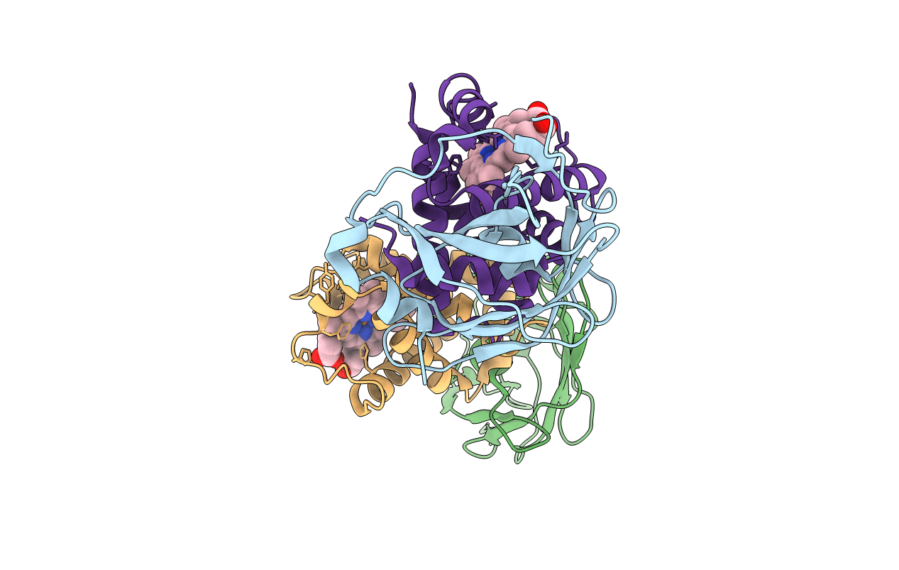
PDB ID:
3S48
Keywords:
Title:
Human Alpha-Haemoglobin Complexed with the First NEAT Domain of IsdH from Staphylococcus aureus
Biological Source:
Source Organism(s):
Staphylococcus aureus (Taxon ID: 282459)
Homo sapiens (Taxon ID: 9606)
Homo sapiens (Taxon ID: 9606)
Expression System(s):
Method Details:
Experimental Method:
Resolution:
3.05 Å
R-Value Free:
0.26
R-Value Work:
0.24
R-Value Observed:
0.24
Space Group:
I 41
