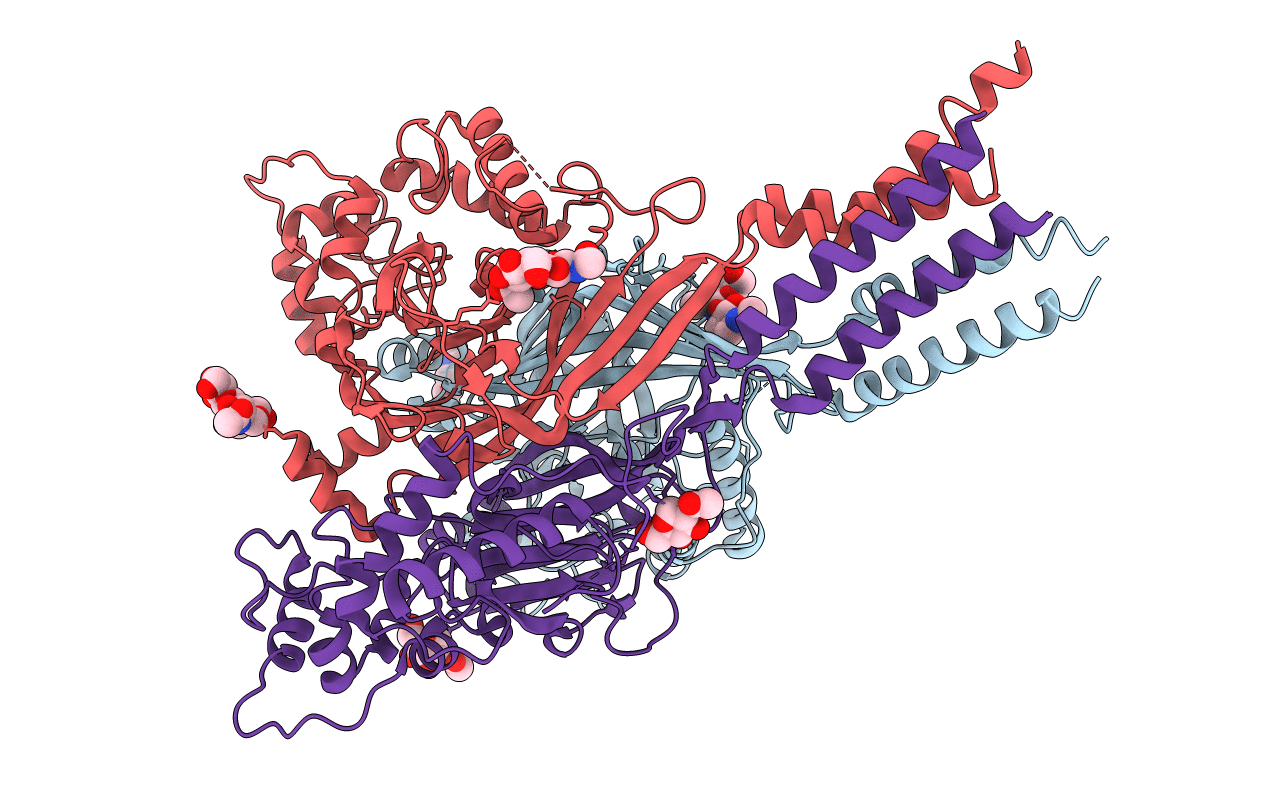
3S3W image
Entry Detail
PDB ID:
3S3W
Title:
Structure of chicken acid-sensing ion channel 1 at 2.6 a resolution and ph 7.5
Biological Source:
Source Organism:
Host Organism:
PDB Version:
Deposition Date:
2011-05-18
Release Date:
2012-05-23
Method Details:
Experimental Method:
Resolution:
2.60 Å
R-Value Free:
0.23
R-Value Work:
0.21
R-Value Observed:
0.21
Space Group:
P 21 21 21
Macromolecular Entities
Polymer Type:polypeptide(L)
Description:Amiloride-sensitive cation channel 2, neuronal
Chain IDs:A, B, C
Chain Length:459
Number of Molecules:3
Biological Source:Gallus gallus
Modified Residue
Compound ID Chain ID Parent Comp ID Details 2D Image
ASN B ASN GLYCOSYLATION SITE
Primary Citation
Structure of the Acid-sensing ion channel 1 in complex with the gating modifier Psalmotoxin 1.
Nat Commun 3 936 936 (2012)
PMID: 22760635 DOI: 10.1038/ncomms1917

Abstact

Venom-derived peptide toxins can modify the gating characteristics of excitatory channels in neurons. How they bind and interfere with the flow of ions without directly blocking the ion permeation pathway remains elusive. Here we report the crystal structure of the trimeric chicken Acid-sensing ion channel 1 in complex with the highly selective gating modifier Psalmotoxin 1 at 3.0 Å resolution. The structure reveals the molecular interactions of three toxin molecules binding at the proton-sensitive acidic pockets of Acid-sensing ion channel 1 and electron density consistent with a cation trapped in the central vestibule above the ion pathway. A hydrophobic patch and a basic cluster are the key structural elements of Psalmotoxin 1 binding, locking two separate regulatory regions in their relative, desensitized-like arrangement. Our results provide a general concept for gating modifier toxin binding suggesting that both surface motifs are required to modify the gating characteristics of an ion channel.

Legend

Protein

Chemical

Disease

Primary Citation of related structures