3S3K image
Deposition Date 2011-05-18
Release Date 2011-11-02
Last Version Date 2023-11-01
Entry Detail
PDB ID:
3S3K
Title:
Crystal structure of the catalytic domain of PTP10D from Drosophila melanogaster with a small molecular inhibitor para-NitroCatechol Sulphate
Biological Source:
Source Organism:
Host Organism:
Method Details:
Experimental Method:
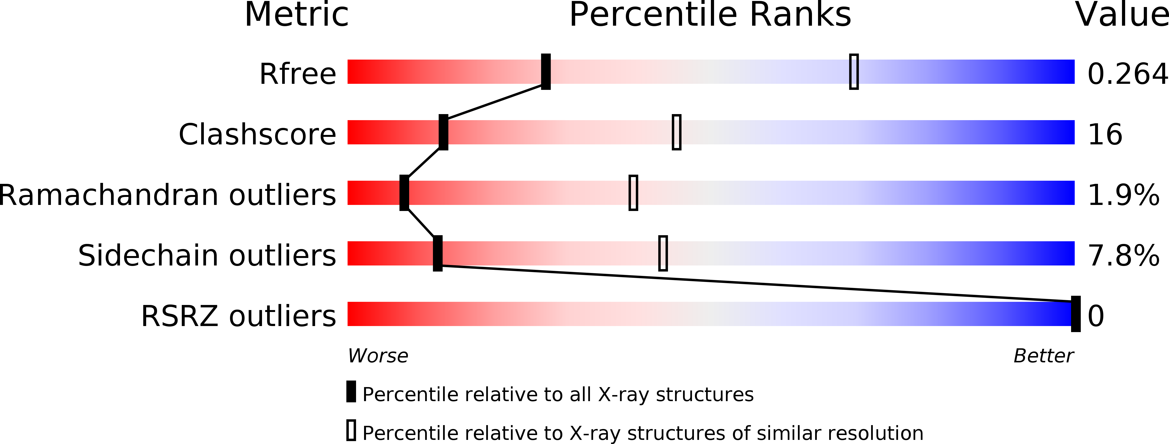
Resolution:
3.20 Å
R-Value Free:
0.30
R-Value Work:
0.25
R-Value Observed:
0.25
Space Group:
P 31 2 1
Macromolecular Entities
Structures with similar UniProt ID
Protein Blast
Polymer Type:polypeptide(L)
Molecule:Tyrosine-protein phosphatase 10D
Gene (Uniprot):Ptp10D
Chain IDs:A, B
Chain Length:307
Number of Molecules:2
Biological Source:Drosophila melanogaster
Ligand Molecules
Primary Citation
Conformational basis for substrate recruitment in protein tyrosine phosphatase 10D
Biochemistry 50 10114 10125 (2011)
PMID: 22007620 DOI: 10.1021/bi201092q

Abstact

The coordinated activity of protein tyrosine phosphatases (PTPs) is crucial for the initiation, modulation, and termination of diverse cellular processes. The catalytic activity of this protein depends on a nucleophilic cysteine at the active site that mediates the hydrolysis of the incoming phosphotyrosine substrate. While the role of conserved residues in the catalytic mechanism of PTPs has been extensively examined, the diversity in the mechanisms of substrate recognition and modulation of catalytic activity suggests that other, less conserved sequence and structural features could contribute to this process. Here we describe the crystal structures of Drosophila melanogaster PTP10D in the apo form as well as in a complex with a substrate peptide and an inhibitor. These studies reveal the role of aromatic ring stacking interactions at the boundary of the active site of PTPs in mediating substrate recruitment. We note that phenylalanine 76, of the so-called KNRY loop, is crucial for orienting the phosphotyrosine residue toward the nucleophilic cysteine. Mutation of phenylalanine 76 to leucine results in a 60-fold decrease in the catalytic efficiency of the enzyme. Fluorescence measurements with a competitive inhibitor, p-nitrocatechol sulfate, suggest that Phe76 also influences the formation of the enzyme-substrate intermediate. The structural and biochemical data for PTP10D thus highlight the role of relatively less conserved residues in PTP domains in both substrate recruitment and modulation of reaction kinetics.

Legend

Protein

Chemical

Disease

Primary Citation of related structures