3S2V image
Deposition Date 2011-05-17
Release Date 2011-06-22
Last Version Date 2024-11-20
Entry Detail
PDB ID:
3S2V
Title:
Crystal Structure of the Ligand Binding Domain of GluK1 in Complex with an Antagonist (S)-1-(2'-Amino-2'-carboxyethyl)-3-[(2-carboxythien-3-yl)methyl]thieno[3,4-d]pyrimidin-2,4-dione at 2.5 A Resolution
Biological Source:
Source Organism:
Host Organism:
Method Details:
Experimental Method:
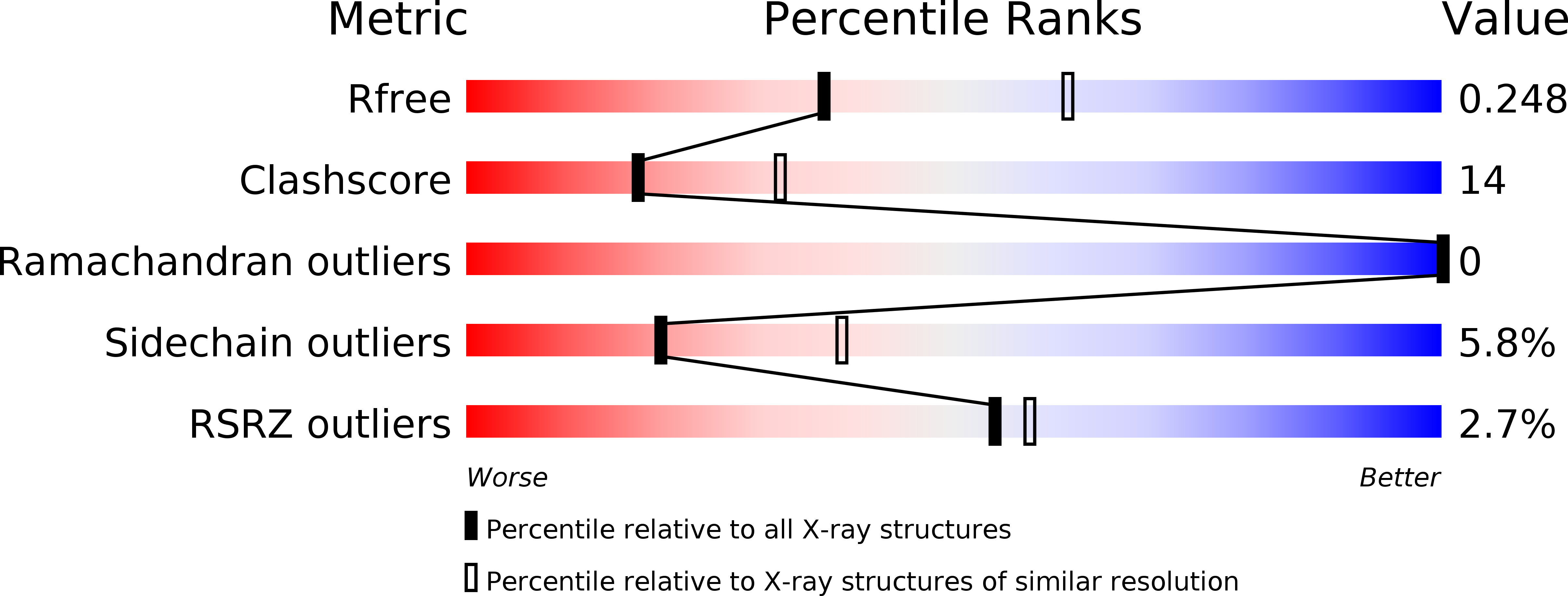
Resolution:
2.50 Å
R-Value Free:
0.25
R-Value Work:
0.17
R-Value Observed:
0.18
Space Group:
P 21 21 21
Macromolecular Entities
Structures with similar UniProt ID
Protein Blast
Polymer Type:polypeptide(L)
Molecule:Glutamate receptor, ionotropic kainate 1
Gene (Uniprot):Grik1
Chain IDs:A, B
Chain Length:257
Number of Molecules:2
Biological Source:Rattus norvegicus
Primary Citation
Selective kainate receptor (GluK1) ligands structurally based upon 1H-cyclopentapyrimidin-2,4(1H,3H)-dione: synthesis, molecular modeling, and pharmacological and biostructural characterization.
J.Med.Chem. 54 4793 4805 (2011)
PMID: 21619066 DOI: 10.1021/jm2004078

Abstact

The physiological function of kainate receptors (GluK1-GluK5) in the central nervous system is not fully understood yet. With the aim of developing potent and selective GluK1 ligands, we have synthesized a series of new thiophene-based GluK1 agonists (6a-c) and antagonists (7a-d). Pharmacological evaluation revealed that they are selective for the GluK1 subunit, with 7b being the most subtype-selective ligand reported to date (GluK1 vs GluK3). The antagonist 7a was cocrystallized with the GluK1 ligand binding domain, and an X-ray crystallographic analysis revealed the largest flexibility in GluK1 ligand binding domain opening upon binding of a ligand seen to date. The results provide new insights into the molecular mechanism of GluK1 receptor ligand binding and pave the way to the development of new tool compounds for studying kainate receptor function.

Legend

Protein

Chemical

Disease

Primary Citation of related structures
Feedback Form
Name
Email
Institute
Feedback