3S2C image
Deposition Date 2011-05-16
Release Date 2012-02-01
Last Version Date 2023-09-13
Entry Detail
PDB ID:
3S2C
Keywords:
Title:
Structure of the thermostable GH51 alpha-L-arabinofuranosidase from Thermotoga petrophila RKU-1
Biological Source:
Source Organism:
Host Organism:
Method Details:
Experimental Method:
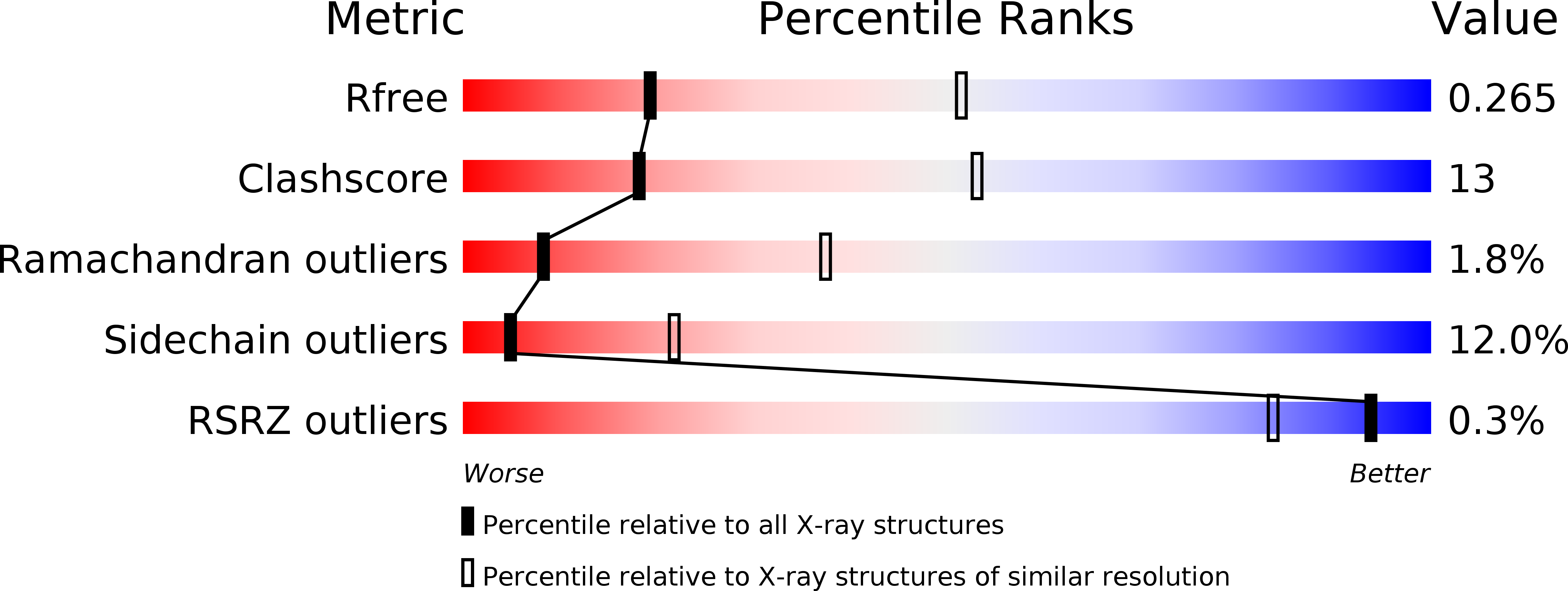
Resolution:
3.00 Å
R-Value Free:
0.26
R-Value Work:
0.19
R-Value Observed:
0.19
Space Group:
P 1 21 1
Macromolecular Entities
Structures with similar UniProt ID
Protein Blast
Polymer Type:polypeptide(L)
Molecule:Alpha-N-arabinofuranosidase
Gene (Uniprot):Tpet_0631
Chain IDs:A, B, C, D, E, F, G, H, I, J, K, L
Chain Length:484
Number of Molecules:12
Biological Source:Thermotoga petrophila
Primary Citation
Structure of a novel thermostable GH51 Alpha-L-arabinofuranosidase from Thermotoga petrophila RKU-1
Protein Sci. 20 1632 1637 (2011)
PMID: 21796714 DOI: 10.1002/pro.693

Abstact

α-L-arabinofuranosidases (EC 3.2.1.55) participate in the degradation of a variety of L-arabinose-containing polysaccharides and interact synergistically with other hemicellulases in the production of oligosaccharides and bioconversion of lignocellulosic biomass into biofuels. In this work, the structure of a novel thermostable family 51 (GH51) α-L-arabinofuranosidase from Thermotoga petrophila RKU-1 (TpAraF) was determined at 3.1 Å resolution. The TpAraF tertiary structure consists of an (α/β)-barrel catalytic core associated with a C-terminal β-sandwich domain, which is stabilized by hydrophobic contacts. In contrast to other structurally characterized GH51 AraFs, the accessory domain of TpAraF is intimately linked to the active site by a long β-hairpin motif, which modifies the catalytic cavity in shape and volume. Sequence and structural analyses indicate that this motif is unique to Thermotoga AraFs. Small angle X-ray scattering investigation showed that TpAraF assembles as a hexamer in solution and is preserved at the optimum catalytic temperature, 65°C, suggesting functional significance. Crystal packing analysis shows that the biological hexamer encompasses a dimer of trimers and the multiple oligomeric interfaces are predominantly fashioned by polar and electrostatic contacts.

Legend

Protein

Chemical

Disease

Primary Citation of related structures
Feedback Form
Name
Email
Institute
Feedback