Deposition Date
2011-05-16
Release Date
2012-06-13
Last Version Date
2023-09-13
Entry Detail
PDB ID:
3S1T
Keywords:
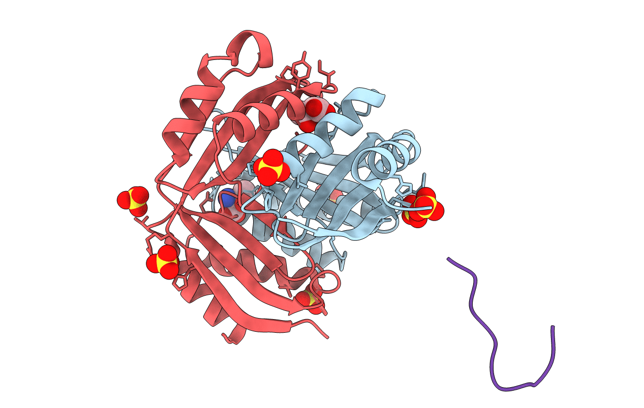
Title:
Structure of the regulatory domain of aspartokinase (Rv3709c; AK-beta) in complex with threonine from Mycobacterium tuberculosis
Biological Source:
Source Organism(s):
Mycobacterium tuberculosis (Taxon ID: 83332)
Expression System(s):
Method Details:
Experimental Method:
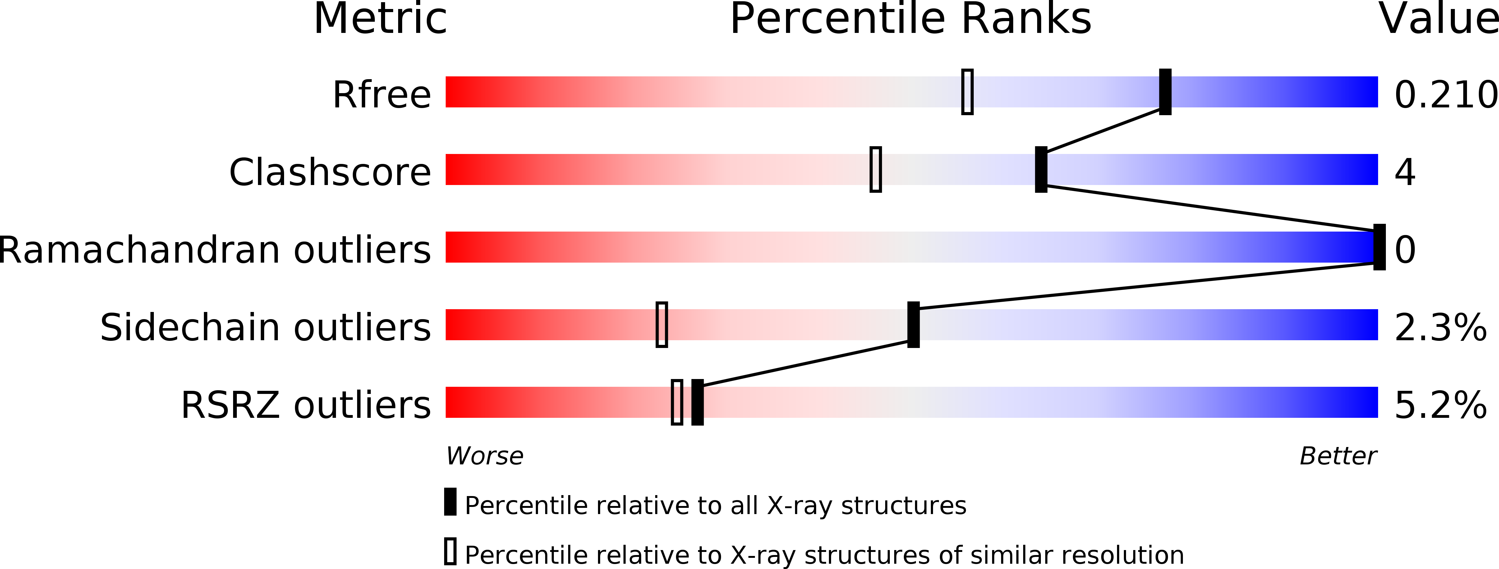
Resolution:
1.63 Å
R-Value Free:
0.21
R-Value Work:
0.19
R-Value Observed:
0.19
Space Group:
P 21 21 21