Deposition Date
2011-05-14
Release Date
2011-09-21
Last Version Date
2024-10-09
Entry Detail
PDB ID:
3S1A
Keywords:
Title:
Crystal structure of the phosphorylation-site double mutant S431E/T432E of the KaiC circadian clock protein
Biological Source:
Source Organism:
Synechococcus elongatus (Taxon ID: 1140)
Host Organism:
Method Details:
Experimental Method:
Resolution:
3.00 Å
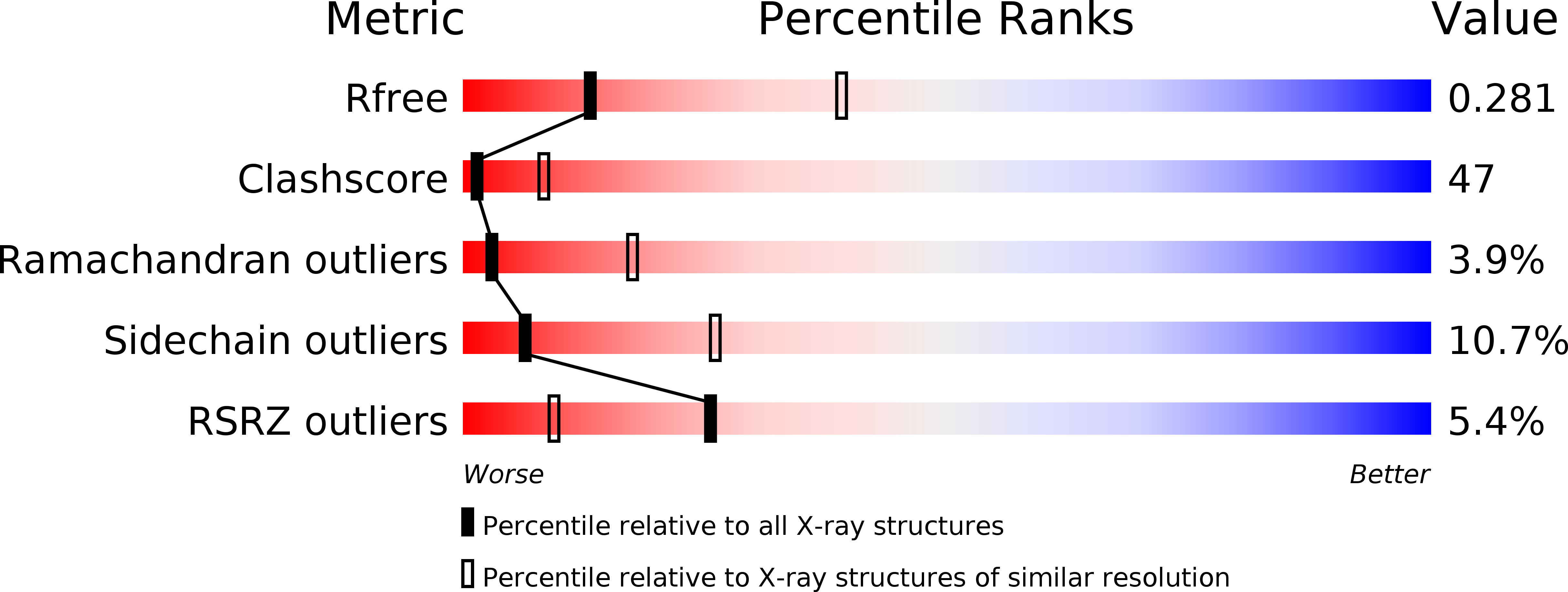
R-Value Free:
0.28
R-Value Work:
0.24
R-Value Observed:
0.26
Space Group:
P 21 21 21