3S02 image
Deposition Date 2011-05-12
Release Date 2012-03-14
Last Version Date 2024-02-28
Entry Detail
PDB ID:
3S02
Keywords:
Title:
The crystal structure of the periplasmic domain of Helicobacter pylori MotB (residues 103-256)
Biological Source:
Source Organism:
Host Organism:
Method Details:
Experimental Method:
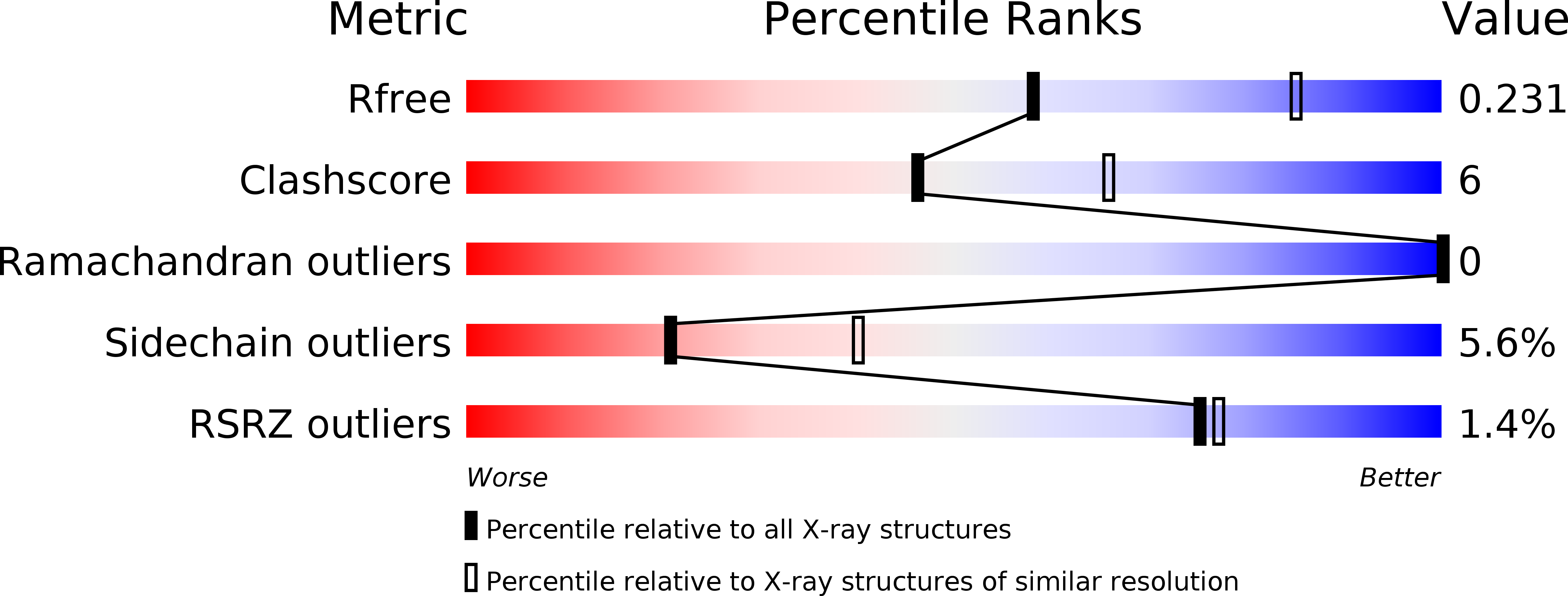
Resolution:
2.50 Å
R-Value Free:
0.24
R-Value Work:
0.18
R-Value Observed:
0.18
Space Group:
P 31 2 1
Macromolecular Entities
Structures with similar UniProt ID
Protein Blast
Polymer Type:polypeptide(L)
Molecule:Motility protein B
Gene (Uniprot):motB
Chain IDs:A, B
Chain Length:154
Number of Molecules:2
Biological Source:Helicobacter pylori
Primary Citation
Role of the MotB linker in the assembly and activation of the bacterial flagellar motor.
Acta Crystallogr.,Sect.D 67 1009 1016 (2011)
PMID: 22120737 DOI: 10.1107/S0907444911041102

Abstact

Bacterial flagella are driven by an ion influx through the peptidoglycan (PG)-tethered MotA/MotB stator. Stator precomplexes assemble in the membrane and remain inactive until they incorporate into the motor, upon which MotA/MotB changes conformation. The nature of this change and the mechanism of inhibition of the PG-binding and ion-conducting activities of the precomplexes are unknown. Here, the structural analysis of a series of N-terminally truncated MotB fragments is presented, the mechanism of inhibition by the linker is identified and the structural basis for the formation of the PG-binding-competent open-channel MotA/MotB conformation via a mechanism that entails linker unfolding and rotational displacement of MotB transmembrane helices is uncovered.

Legend

Protein

Chemical

Disease

Primary Citation of related structures
Feedback Form
Name
Email
Institute
Feedback