3RZV image
Deposition Date 2011-05-12
Release Date 2011-10-19
Last Version Date 2024-11-27
Entry Detail
PDB ID:
3RZV
Keywords:
Title:
The Crystal Structure of a E280A Mutant of the Catalytic Domain of AMSH
Biological Source:
Source Organism:
Homo sapiens (Taxon ID: 9606)
Host Organism:
Method Details:
Experimental Method:
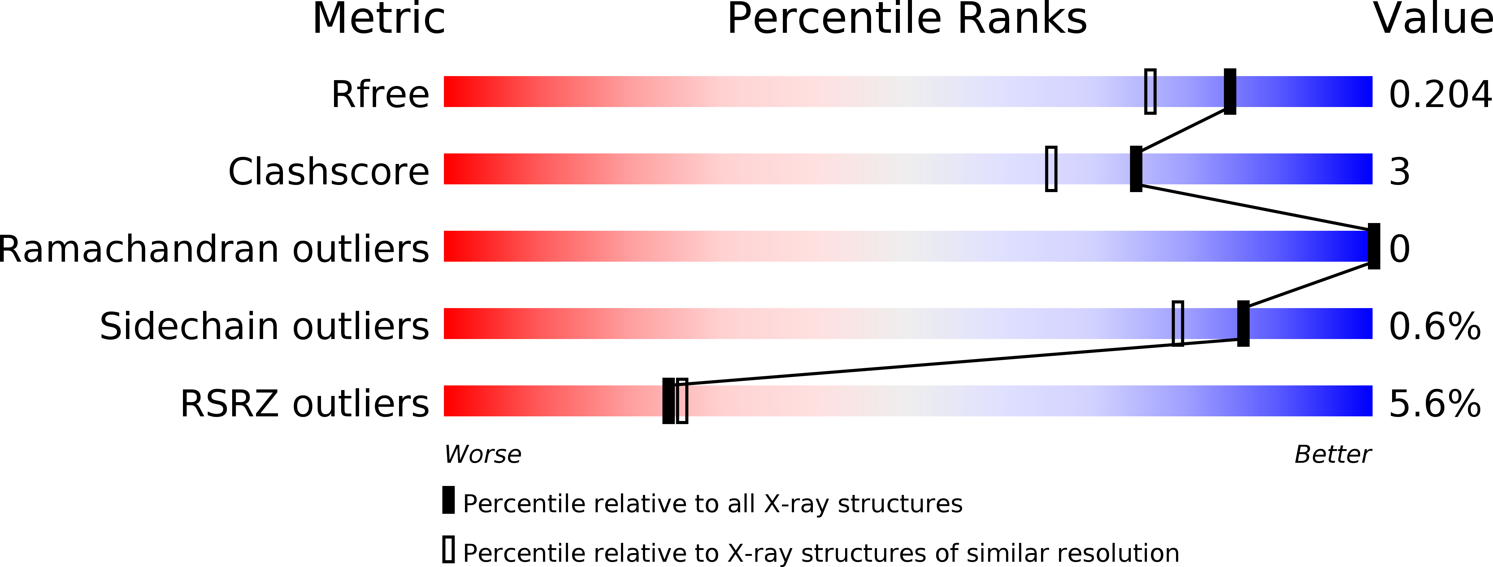
Resolution:
1.67 Å
R-Value Free:
0.20
R-Value Work:
0.18
R-Value Observed:
0.18
Space Group:
P 43 21 2
Macromolecular Entities
Polymer Type:polypeptide(L)
Molecule:STAM-binding protein
Gene (Uniprot):STAMBP
Mutations:E280A
Chain IDs:A
Chain Length:211
Number of Molecules:1
Biological Source:Homo sapiens
Ligand Molecules
Primary Citation
Structural and Thermodynamic Comparison of the Catalytic Domain of AMSH and AMSH-LP: Nearly Identical Fold but Different Stability.
J.Mol.Biol. 413 416 429 (2011)
PMID: 21888914 DOI: 10.1016/j.jmb.2011.08.029

Abstact

AMSH plays a critical role in the ESCRT (endosomal sorting complexes required for transport) machinery, which facilitates the down-regulation and degradation of cell-surface receptors. It displays a high level of specificity toward cleavage of Lys63-linked polyubiquitin chains, the structural basis of which has been understood recently through the crystal structure of a highly related, but ESCRT-independent, protein AMSH-LP (AMSH-like protein). We have determined the X-ray structure of two constructs representing the catalytic domain of AMSH: AMSH244, the JAMM (JAB1/MPN/MOV34)-domain-containing polypeptide segment from residues 244 to 424, and AMSH219(E280A), an active-site mutant, Glu280 to Ala, of the segment from 219 to 424. In addition to confirming the expected zinc coordination in the protein, the structures reveal that the catalytic domains of AMSH and AMSH-LP are nearly identical; however, guanidine-hydrochloride-induced unfolding studies show that the catalytic domain of AMSH is thermodynamically less stable than that of AMSH-LP, indicating that the former is perhaps structurally more plastic. Much to our surprise, in the AMSH219(E280A) structure, the catalytic zinc was still held in place, by the compensatory effect of an aspartate from a nearby loop moving into a position where it could coordinate with the zinc, once again suggesting the plasticity of AMSH. Additionally, a model of AMSH244 bound to Lys63-linked diubiquitin reveals a type of interface for the distal ubiquitin significantly different from that seen in AMSH-LP. Altogether, we believe that our data provide important insight into the structural difference between the two proteins that may translate into the difference in their biological function.

Legend

Protein

Chemical

Disease

Primary Citation of related structures