3RZC image
Deposition Date 2011-05-11
Release Date 2011-08-24
Last Version Date 2024-11-20
Entry Detail
PDB ID:
3RZC
Keywords:
Title:
Structure of the self-antigen iGb3 bound to mouse CD1d and in complex with the iNKT TCR
Biological Source:
Source Organism:
Host Organism:
Method Details:
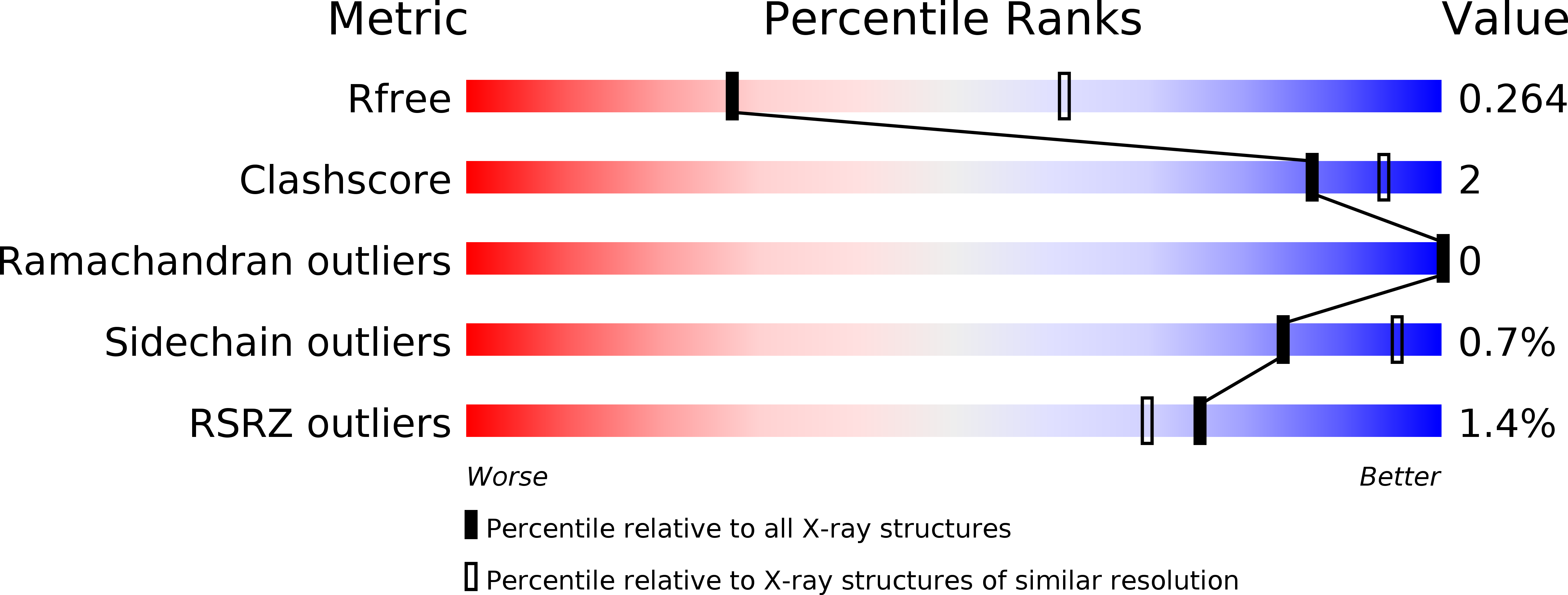
Experimental Method:
Resolution:
2.80 Å
R-Value Free:
0.27
R-Value Work:
0.23
R-Value Observed:
0.23
Space Group:
C 2 2 21
Macromolecular Entities
Structures with similar UniProt ID
Protein Blast
Polymer Type:polypeptide(L)
Molecule:Antigen-presenting glycoprotein CD1d1
Gene (Uniprot):Cd1d1
Chain IDs:A
Chain Length:285
Number of Molecules:1
Biological Source:Mus musculus
Structures with similar UniProt ID
Protein Blast
Polymer Type:polypeptide(L)
Molecule:Beta-2-microglobulin
Gene (Uniprot):B2m
Chain IDs:B
Chain Length:99
Number of Molecules:1
Biological Source:Mus musculus
Polymer Type:polypeptide(L)
Molecule:Valpha14
Chain IDs:C
Chain Length:209
Number of Molecules:1
Biological Source:Mus musculus, Homo sapiens
Polymer Type:polypeptide(L)
Molecule:Vbeta8.2
Chain IDs:D
Chain Length:241
Number of Molecules:1
Biological Source:Mus musculus, Homo sapiens
Modified Residue
Compound ID Chain ID Parent Comp ID Details 2D Image
ASN A ASN GLYCOSYLATION SITE
Ligand Molecules
Primary Citation
Cutting Edge: Structural Basis for the Recognition of {beta}-Linked Glycolipid Antigens by Invariant NKT Cells.
J.Immunol. 187 2079 2083 (2011)
PMID: 21810611 DOI: 10.4049/jimmunol.1101636

Abstact

Invariant NKT (iNKT) cells expressing a semi-invariant Vα14 TCR recognize self and foreign lipid Ags when presented by the nonclassical MHCI homolog CD1d. Whereas the majority of known iNKT cell Ags are characterized by the presence of a single α-linked sugar, mammalian self Ags are β-linked glycosphingolipids, posing the interesting question of how the semi-invariant TCR can bind to such structurally distinct ligands. In this study, we show that the mouse iNKT TCR recognizes the complex β-linked Ag isoglobotrihexosylceramide (iGb3; Galα1-3-Galβ1-4-Glcβ1-1Cer) by forcing the proximal β-linked sugar of the trisaccharide head group to adopt the typical binding orientation of α-linked glycolipids. The squashed iGb3 orientation is stabilized by several interactions between the trisaccharide and CD1d residues. Finally, the formation of novel contacts between the proximal and second sugar of iGb3 and CDR2α residues of the TCR suggests an expanded recognition logic that can possibly distinguish foreign Ags from self Ags.

Legend

Protein

Chemical

Disease

Primary Citation of related structures
Feedback Form
Name
Email
Institute
Feedback