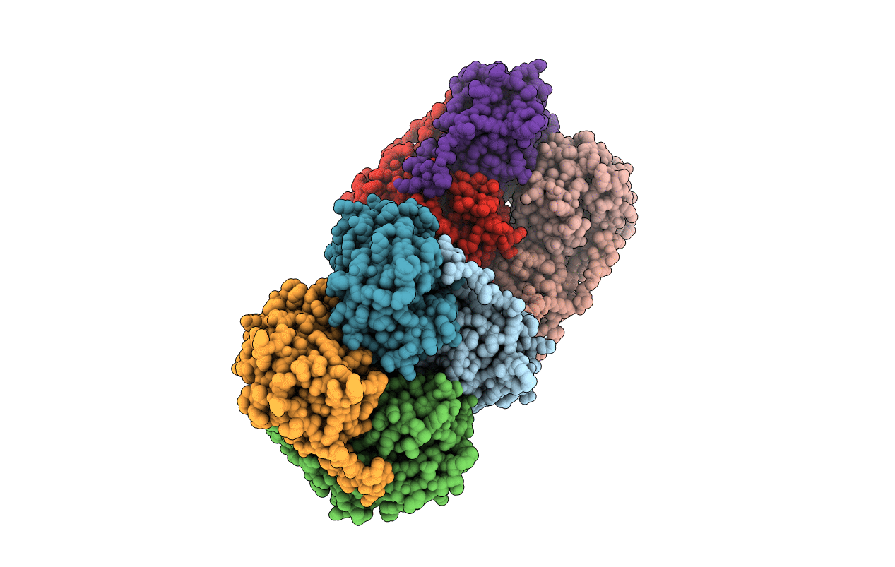
3RWA image
Entry Detail
PDB ID:
3RWA
Title:
Crystal structure of circular-permutated mKate
Biological Source:
Source Organism:
Host Organism:
PDB Version:
Deposition Date:
2011-05-08
Release Date:
2011-06-15
Method Details:
Experimental Method:
Resolution:
1.67 Å
R-Value Free:
0.21
R-Value Work:
0.18
R-Value Observed:
0.18
Space Group:
C 1 2 1
Macromolecular Entities
Polymer Type:polypeptide(L)
Description:Fluorescent protein FP480
Chain IDs:A, B, C, D, E, F, G, H
Chain Length:235
Number of Molecules:8
Biological Source:Entacmaea quadricolor
Modified Residue
Compound ID Chain ID Parent Comp ID Details 2D Image
MET A GLN chromophore
NRQ A GLY chromophore
Primary Citation
Circular permutation of red fluorescent proteins.
Plos One 6 e20505 e20505 (2011)
PMID: 21647365 DOI: 10.1371/journal.pone.0020505

Abstact

Circular permutation of fluorescent proteins provides a substrate for the design of molecular sensors. Here we describe a systematic exploration of permutation sites for mCherry and mKate using a tandem fusion template approach. Circular permutants retaining more than 60% (mCherry) and 90% (mKate) brightness of the parent molecules are reported, as well as a quantitative evaluation of the fluorescence from neighboring mutations. Truncations of circular permutants indicated essential N- and C-terminal segments and substantial flexibility in the use of these molecules. Structural evaluation of two cp-mKate variants indicated no major conformational changes from the previously reported wild-type structure, and cis conformation of the chromophores. Four cp-mKates were identified with over 80% of native fluorescence, providing important new building blocks for sensor and complementation experiments.

Legend

Protein

Chemical

Disease

Primary Citation of related structures