Deposition Date
2011-05-06
Release Date
2012-05-09
Last Version Date
2023-09-13
Entry Detail
PDB ID:
3RVM
Keywords:
Title:
Structure of the CheY-Mn2+ Complex with substitutions at 59 and 89: N59D and E89R
Biological Source:
Source Organism(s):
Escherichia coli (Taxon ID: 83333)
Expression System(s):
Method Details:
Experimental Method:
Resolution:
1.45 Å
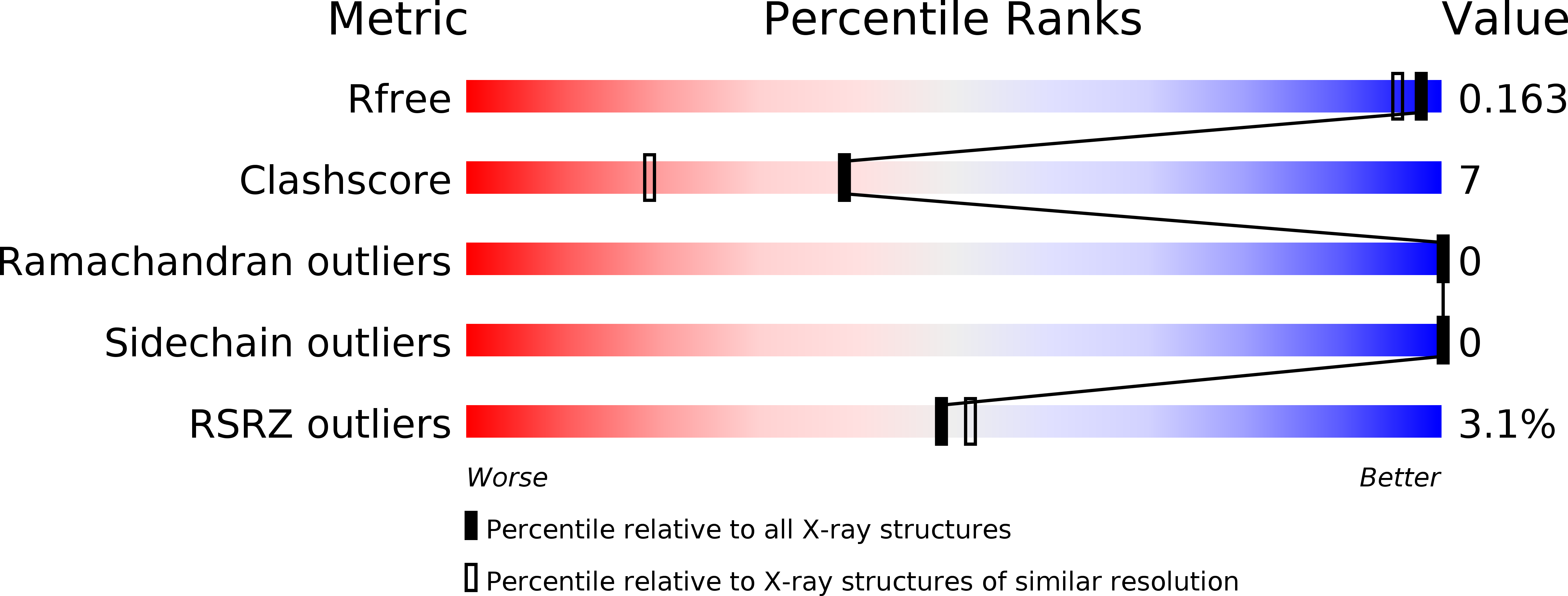
R-Value Free:
0.16
R-Value Work:
0.13
R-Value Observed:
0.14
Space Group:
P 21 21 21