3RV5 image
Deposition Date 2011-05-06
Release Date 2011-08-31
Last Version Date 2024-10-16
Entry Detail
PDB ID:
3RV5
Title:
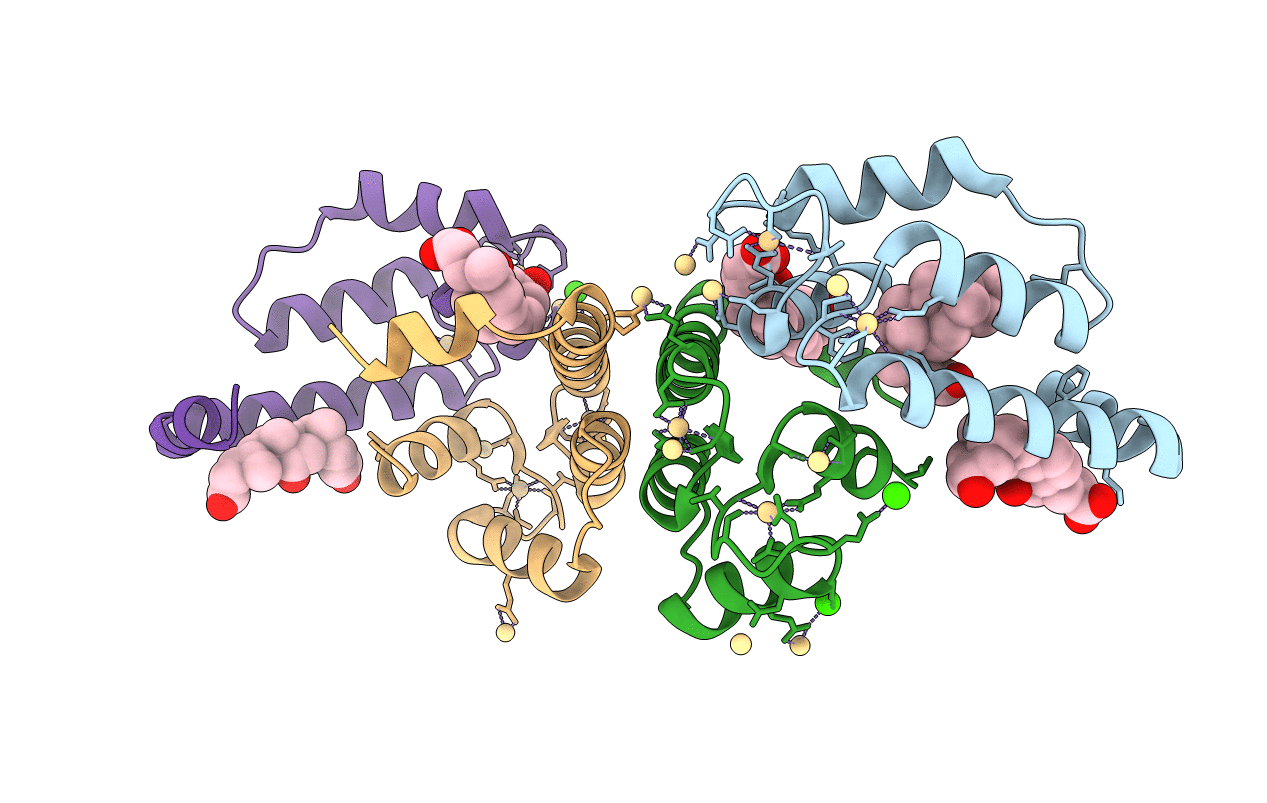
Crystal structure of human cardiac troponin C regulatory domain in complex with cadmium and deoxycholic acid
Biological Source:
Source Organism:
Homo sapiens (Taxon ID: 9606)
Host Organism:
Method Details:
Experimental Method:
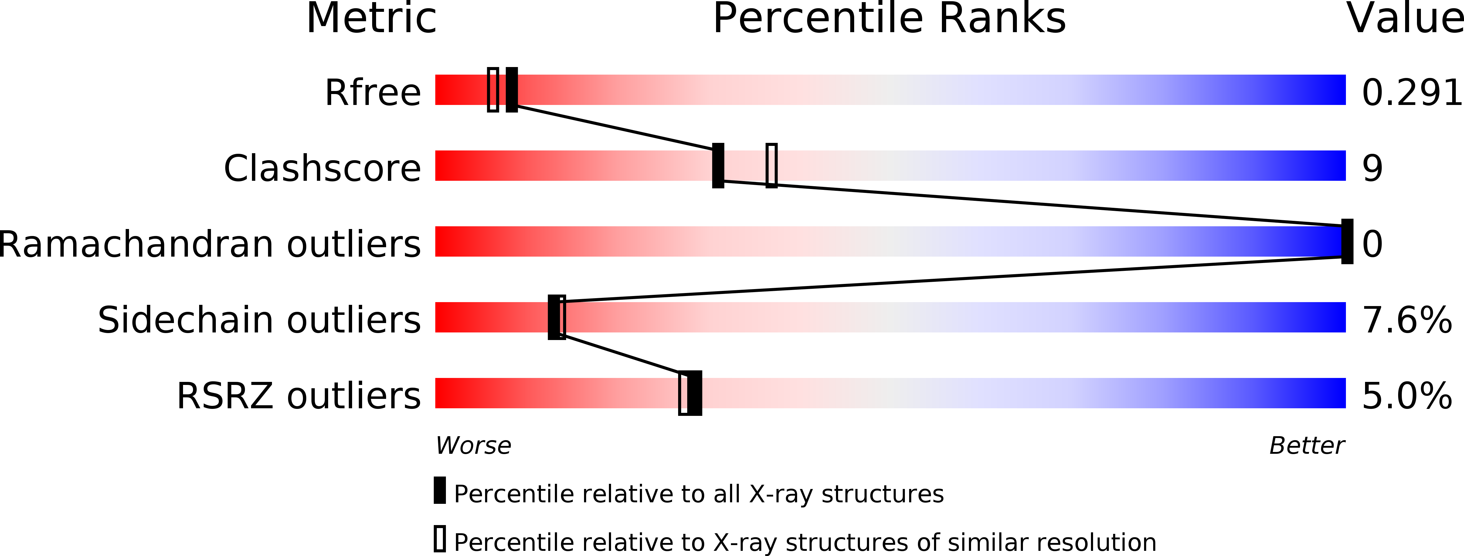
Resolution:
2.20 Å
R-Value Free:
0.28
R-Value Work:
0.22
R-Value Observed:
0.22
Space Group:
P 21 21 2
Macromolecular Entities
Structures with similar UniProt ID
Protein Blast
Polymer Type:polypeptide(L)
Molecule:Troponin C, slow skeletal and cardiac muscles
Gene (Uniprot):TNNC1
Chain IDs:A, B, C, D
Chain Length:89
Number of Molecules:4
Biological Source:Homo sapiens
Modified Residue
Compound ID Chain ID Parent Comp ID Details 2D Image
MSE A MET SELENOMETHIONINE
Primary Citation
Crystal structure of cardiac troponin C regulatory domain in complex with cadmium and deoxycholic Acid reveals novel conformation.
J.Mol.Biol. 413 699 711 (2011)
PMID: 21920370 DOI: 10.1016/j.jmb.2011.08.049

Abstact

The amino-terminal regulatory domain of cardiac troponin C (cNTnC) plays an important role as the calcium sensor for the troponin complex. Calcium binding to cNTnC results in conformational changes that trigger a cascade of events that lead to cardiac muscle contraction. The cardiac N-terminal domain of TnC consists of two EF-hand calcium binding motifs, one of which is dysfunctional in binding calcium. Nevertheless, the defunct EF-hand still maintains a role in cNTnC function. For its structural analysis by X-ray crystallography, human cNTnC with the wild-type primary sequence was crystallized under a novel crystallization condition. The crystal structure was solved by the single-wavelength anomalous dispersion method and refined to 2.2 Å resolution. The structure displays several novel features. Firstly, both EF-hand motifs coordinate cadmium ions derived from the crystallization milieu. Secondly, the ion coordination in the defunct EF-hand motif accompanies unusual changes in the protein conformation. Thirdly, deoxycholic acid, also derived from the crystallization milieu, is bound in the central hydrophobic cavity. This is reminiscent of the interactions observed for cardiac calcium sensitizer drugs that bind to the same core region and maintain the "open" conformational state of calcium-bound cNTnC. The cadmium ion coordination in the defunct EF-hand indicates that this vestigial calcium binding site retains the structural and functional elements that allow it to coordinate a cadmium ion. However, it is a result of, or concomitant with, large and unusual structural changes in cNTnC.

Legend

Protein

Chemical

Disease

Primary Citation of related structures