Deposition Date
2011-05-05
Release Date
2011-08-03
Last Version Date
2024-02-28
Entry Detail
PDB ID:
3RV0
Keywords:
Title:
Crystal structure of K. polysporus Dcr1 without the C-terminal dsRBD
Biological Source:
Source Organism(s):
Vanderwaltozyma polyspora (Taxon ID: 436907)
Expression System(s):
Method Details:
Experimental Method:
Resolution:
2.29 Å
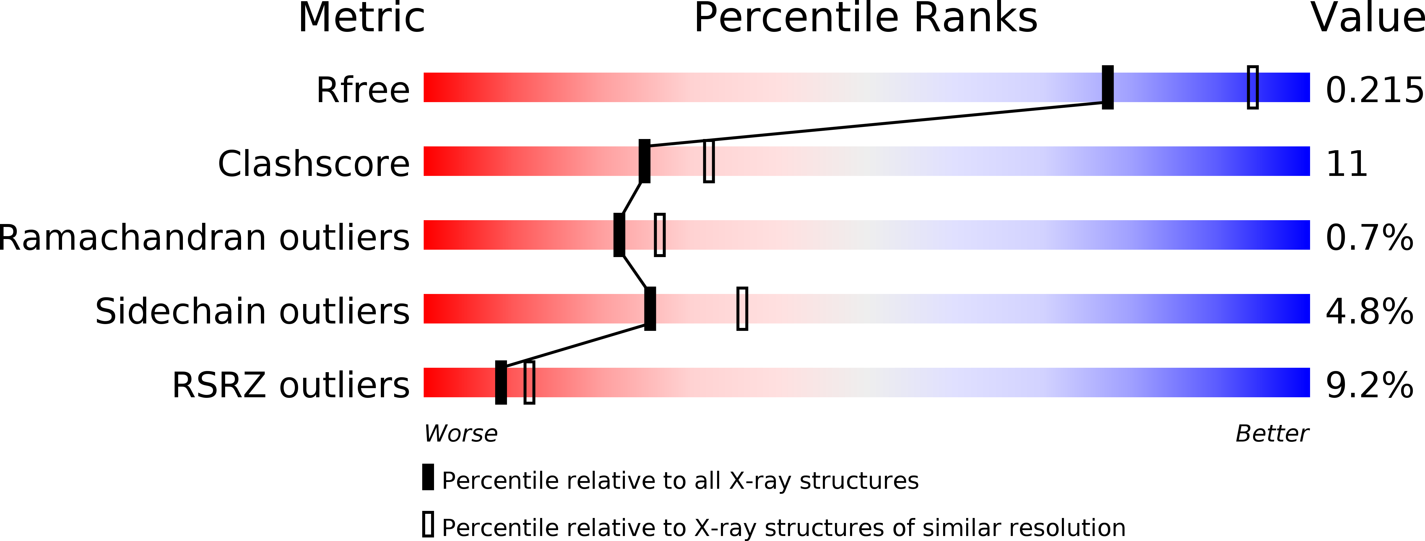
R-Value Free:
0.21
R-Value Work:
0.17
R-Value Observed:
0.17
Space Group:
P 21 21 21