
Deposition Date
2011-04-29
Release Date
2011-08-10
Last Version Date
2024-04-03
Entry Detail
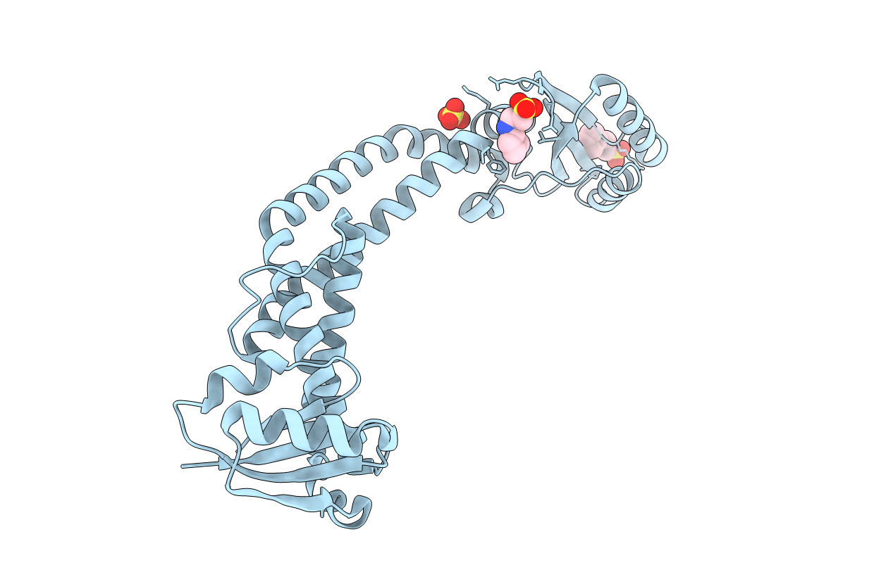
PDB ID:
3RRK
Keywords:
Title:
Crystal structure of the cytoplasmic N-terminal domain of subunit I, homolog of subunit a, of V-ATPase
Biological Source:
Source Organism(s):
Meiothermus ruber (Taxon ID: 504728)
Expression System(s):
Method Details:
Experimental Method:
Resolution:
2.64 Å
R-Value Free:
0.25
R-Value Work:
0.18
R-Value Observed:
0.19
Space Group:
I 2 2 2
