3RQF image
Deposition Date 2011-04-28
Release Date 2011-06-01
Last Version Date 2023-09-13
Entry Detail
PDB ID:
3RQF
Keywords:
Title:
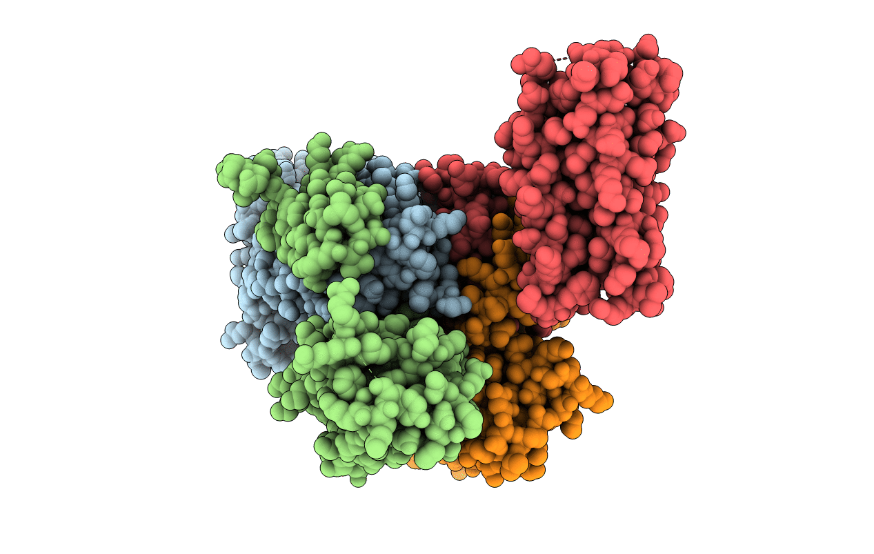
Cerebral cavernous malformation 3 (CCM3) in complex with paxillin LD2
Biological Source:
Source Organism:
Homo sapiens (Taxon ID: 9606)
Host Organism:
Method Details:
Experimental Method:
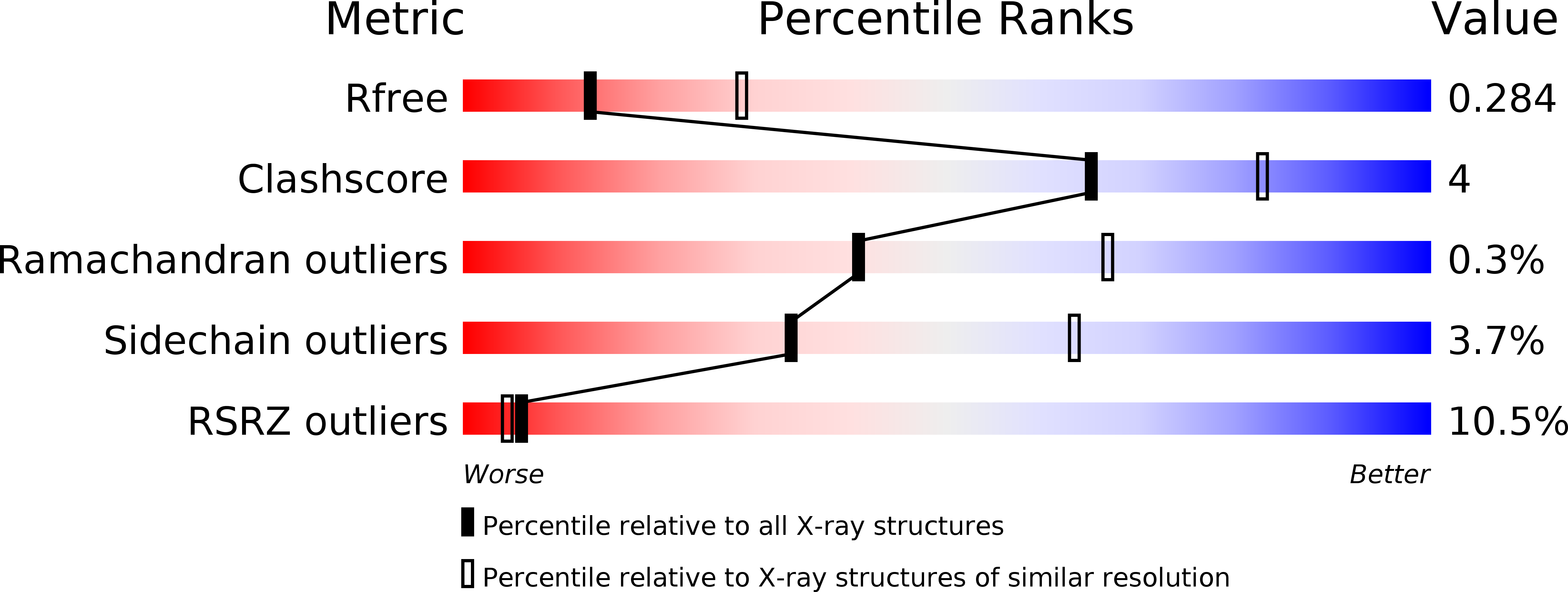
Resolution:
2.70 Å
R-Value Free:
0.29
R-Value Work:
0.23
R-Value Observed:
0.23
Space Group:
P 21 21 21
Macromolecular Entities
Structures with similar UniProt ID
Protein Blast
Polymer Type:polypeptide(L)
Molecule:Programmed cell death protein 10
Gene (Uniprot):PDCD10
Chain IDs:A, B, C, D
Chain Length:214
Number of Molecules:4
Biological Source:Homo sapiens
Structures with similar UniProt ID
Protein Blast
Polymer Type:polypeptide(L)
Molecule:Paxillin LD2 peptide
Chain IDs:E
Chain Length:13
Number of Molecules:1
Biological Source:Homo Sapiens
Primary Citation
Molecular Recognition of Leucine-Aspartate Repeat (LD) Motifs by the Focal Adhesion Targeting Homology Domain of Cerebral Cavernous Malformation 3 (CCM3).
J.Biol.Chem. 286 26138 26147 (2011)
PMID: 21632544 DOI: 10.1074/jbc.M110.211250

Abstact

Cerebral cavernous malformation (CCM) is a disease that affects between 0.1 and 0.5% of the human population, with mutations in CCM3 accounting for ~ 15% of the autosomal dominant form of the disease. We recently reported that CCM3 contains an N-terminal dimerization domain (CCM3D) and a C-terminal focal adhesion targeting (FAT) homology domain. Intermolecular protein-protein interactions of CCM3 are mediated by a highly conserved surface on the FAT homology domain and are affected by CCM3 truncations in the human disease. Here we report the crystal structures of CCM3 in complex with three different leucine-aspartate repeat (LD) motifs (LD1, LD2, and LD4) from the scaffolding protein paxillin, at 2.8, 2.7, and 2.5 Å resolution. We show that CCM3 binds LD motifs using the highly conserved hydrophobic patch 1 (HP1) and that this binding is similar to the binding of focal adhesion kinase and Pyk2 FAT domains to paxillin LD motifs. We further show by surface plasmon resonance that CCM3 binds paxillin LD motifs with affinities in the micromolar range, similar to FAK family FAT domains. Finally, we show that endogenous CCM3 and paxillin co-localize in mouse cerebral pericytes. These studies provide a molecular-level framework to investigate the protein-protein interactions of CCM3.

Legend

Protein

Chemical

Disease

Primary Citation of related structures
Feedback Form
Name
Email
Institute
Feedback