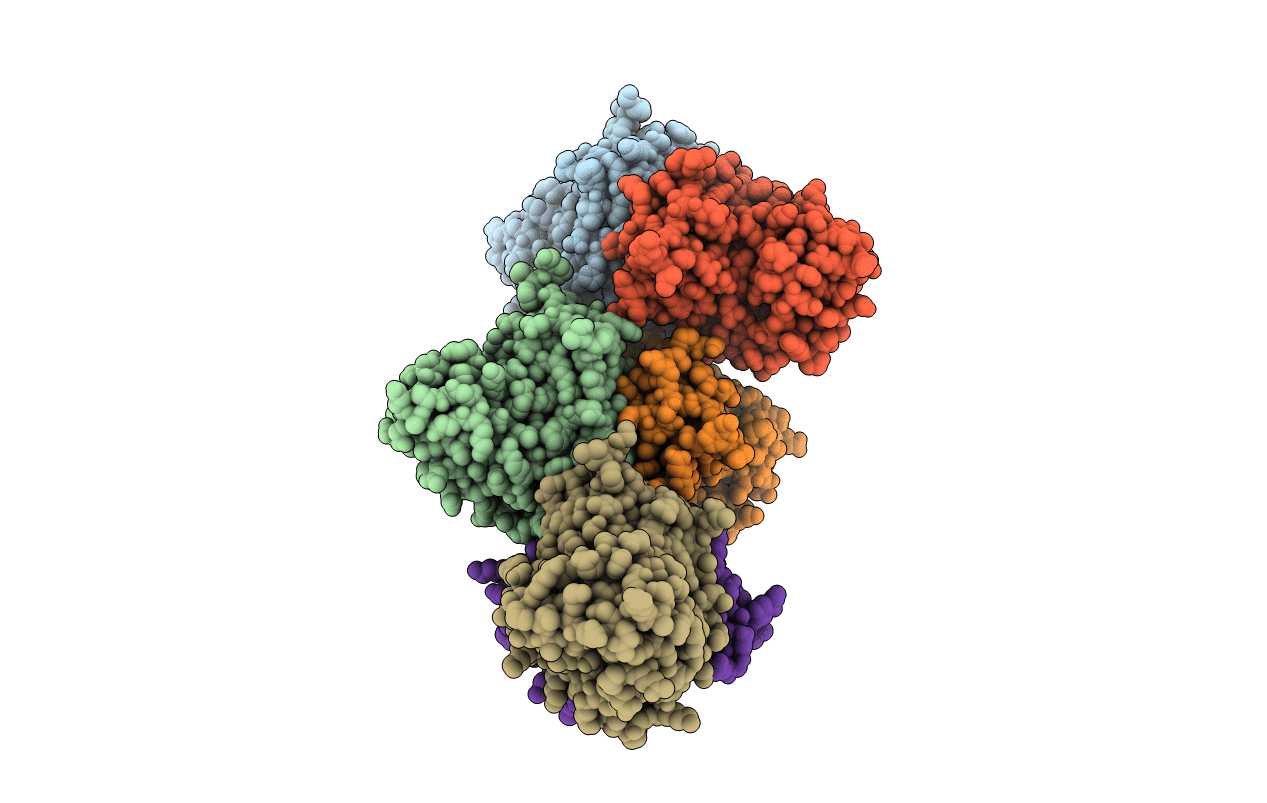
3RPN image
Deposition Date 2011-04-27
Release Date 2011-07-13
Last Version Date 2023-11-01
Entry Detail
PDB ID:
3RPN
Title:
Crystal structure of human kappa class glutathione transferase in complex with S-hexylglutathione
Biological Source:
Source Organism:
Homo sapiens (Taxon ID: 9606)
Host Organism:
Method Details:
Experimental Method:
Resolution:
1.90 Å
R-Value Free:
0.21
R-Value Work:
0.18
R-Value Observed:
0.18
Space Group:
P 1 21 1
Macromolecular Entities
Polymer Type:polypeptide(L)
Molecule:Glutathione S-transferase kappa 1
Gene (Uniprot):GSTK1
Chain IDs:A, B, C, D, E, F
Chain Length:234
Number of Molecules:6
Biological Source:Homo sapiens
Ligand Molecules
Primary Citation
Crystal structures and kinetic studies of human Kappa class glutathione transferase provide insights into the catalytic mechanism.
Biochem.J. 439 215 225 (2011)
PMID: 21728995 DOI: 10.1042/BJ20110753

Abstact

GSTs (glutathione transferases) are a family of enzymes that primarily catalyse nucleophilic addition of the thiol of GSH (reduced glutathione) to a variety of hydrophobic electrophiles in the cellular detoxification of cytotoxic and genotoxic compounds. GSTks (Kappa class GSTs) are a distinct class because of their unique cellular localization, function and structure. In the present paper we report the crystal structures of hGSTk (human GSTk) in apo-form and in complex with GTX (S-hexylglutathione) and steady-state kinetic studies, revealing insights into the catalytic mechanism of hGSTk and other GSTks. Substrate binding induces a conformational change of the active site from an 'open' conformation in the apo-form to a 'closed' conformation in the GTX-bound complex, facilitating formations of the G site (GSH-binding site) and the H site (hydrophobic substrate-binding site). The conserved Ser(16) at the G site functions as the catalytic residue in the deprotonation of the thiol group and the conserved Asp(69), Ser(200), Asp(201) and Arg(202) form a network of interactions with γ-glutamyl carboxylate to stabilize the thiolate anion. The H site is a large hydrophobic pocket with conformational flexibility to allow the binding of different hydrophobic substrates. The kinetic mechanism of hGSTk conforms to a rapid equilibrium random sequential Bi Bi model.

Legend

Protein

Chemical

Disease

Primary Citation of related structures