
Deposition Date
2011-04-27
Release Date
2011-10-26
Last Version Date
2024-11-20
Entry Detail
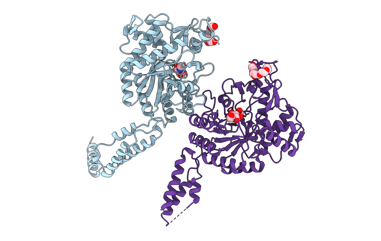
PDB ID:
3RPM
Keywords:
Title:
Crystal structure of the first GH20 domain of a novel Beta-N-acetyl-hexosaminidase StrH from Streptococcus pneumoniae R6
Biological Source:
Source Organism(s):
Streptococcus pneumoniae (Taxon ID: 171101)
Expression System(s):
Method Details:
Experimental Method:
Resolution:
2.10 Å
R-Value Free:
0.22
R-Value Work:
0.19
R-Value Observed:
0.19
Space Group:
P 31 2 1
