3RNM image
Deposition Date 2011-04-22
Release Date 2011-05-04
Last Version Date 2025-03-26
Entry Detail
PDB ID:
3RNM
Title:
The crystal structure of the subunit binding of human dihydrolipoamide transacylase (E2b) bound to human dihydrolipoamide dehydrogenase (E3)
Biological Source:
Source Organism:
Homo sapiens (Taxon ID: 9606)
Host Organism:
Method Details:
Experimental Method:
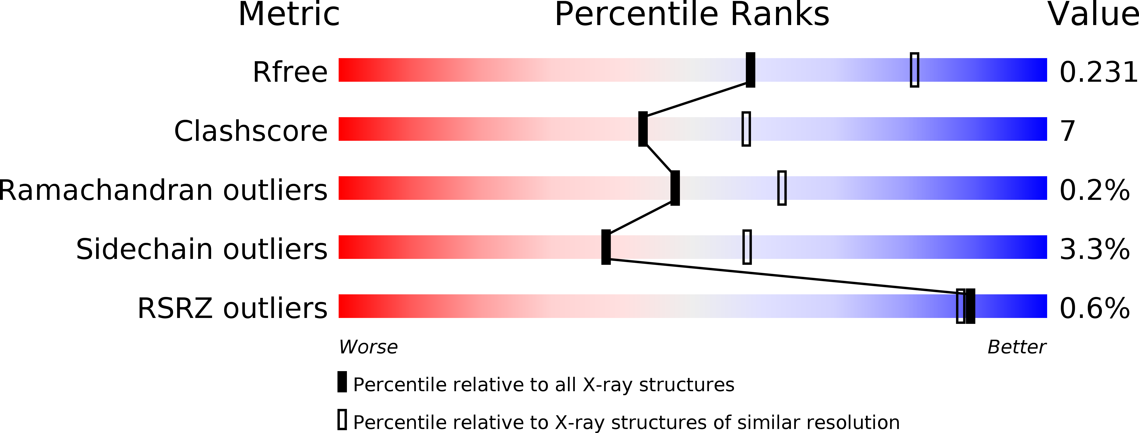
Resolution:
2.40 Å
R-Value Free:
0.23
R-Value Work:
0.18
R-Value Observed:
0.18
Space Group:
P 1 21 1
Macromolecular Entities
Polymer Type:polypeptide(L)
Molecule:Dihydrolipoyl dehydrogenase, mitochondrial
Gene (Uniprot):DLD
Chain IDs:A, B, C, D
Chain Length:495
Number of Molecules:4
Biological Source:Homo sapiens
Polymer Type:polypeptide(L)
Molecule:Lipoamide acyltransferase component of branched-chain alpha-keto acid dehydrogenase complex, mitochondrial
Gene (Uniprot):DBT
Mutations:R118N
Chain IDs:E, F
Chain Length:58
Number of Molecules:2
Biological Source:Homo sapiens
Primary Citation
Structural and Thermodynamic Basis for Weak Interactions between Dihydrolipoamide Dehydrogenase and Subunit-binding Domain of the Branched-chain {alpha}-Ketoacid Dehydrogenase Complex.
J.Biol.Chem. 286 23476 23488 (2011)
PMID: 21543315 DOI: 10.1074/jbc.M110.202960

Abstact

The purified mammalian branched-chain α-ketoacid dehydrogenase complex (BCKDC), which catalyzes the oxidative decarboxylation of branched-chain α-keto acids, is essentially devoid of the constituent dihydrolipoamide dehydrogenase component (E3). The absence of E3 is associated with the low affinity of the subunit-binding domain of human BCKDC (hSBDb) for hE3. In this work, sequence alignments of hSBDb with the E3-binding domain (E3BD) of the mammalian pyruvate dehydrogenase complex show that hSBDb has an arginine at position 118, where E3BD features an asparagine. Substitution of Arg-118 with an asparagine increases the binding affinity of the R118N hSBDb variant (designated hSBDb*) for hE3 by nearly 2 orders of magnitude. The enthalpy of the binding reaction changes from endothermic with the wild-type hSBDb to exothermic with the hSBDb* variant. This higher affinity interaction allowed the determination of the crystal structure of the hE3/hSBDb* complex to 2.4-Å resolution. The structure showed that the presence of Arg-118 poses a unique, possibly steric and/or electrostatic incompatibility that could impede E3 interactions with the wild-type hSBDb. Compared with the E3/E3BD structure, the hE3/hSBDb* structure has a smaller interfacial area. Solution NMR data corroborated the interactions of hE3 with Arg-118 and Asn-118 in wild-type hSBDb and mutant hSBDb*, respectively. The NMR results also showed that the interface between hSBDb and hE3 does not change significantly from hSBDb to hSBDb*. Taken together, our results represent a starting point for explaining the long standing enigma that the E2b core of the BCKDC binds E3 far more weakly relative to other α-ketoacid dehydrogenase complexes.

Legend

Protein

Chemical

Disease

Primary Citation of related structures