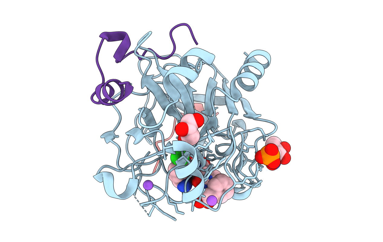
3RML image
Entry Detail
PDB ID:
3RML
Title:
Human Thrombin in complex with MI331
Biological Source:
PDB Version:
Deposition Date:
2011-04-21
Release Date:
2012-04-25
Method Details:
Experimental Method:
Resolution:
1.53 Å
R-Value Free:
0.18
R-Value Work:
0.15
R-Value Observed:
0.16
Space Group:
C 1 2 1
Macromolecular Entities
Polymer Type:polypeptide(L)
Description:Thrombin Heavy Chain
Chain IDs:B (auth: H)
Chain Length:259
Number of Molecules:1
Biological Source:Homo sapiens
Polymer Type:polypeptide(L)
Description:Hirudin variant-2
Chain IDs:C (auth: I)
Chain Length:13
Number of Molecules:1
Biological Source:Hirudo medicinalis
Polymer Type:polypeptide(L)
Description:Thrombin Light Chain
Chain IDs:A (auth: L)
Chain Length:36
Number of Molecules:1
Biological Source:Homo sapiens
Modified Residue
Compound ID Chain ID Parent Comp ID Details 2D Image
ASN B ASN GLYCOSYLATION SITE
TYS C TYR O-SULFO-L-TYROSINE
Peptide-like Molecules
PRD_001090
Primary Citation
Ligand binding stepwise disrupts water network in thrombin: enthalpic and entropic changes reveal classical hydrophobic effect
J.Med.Chem. 55 6094 6110 (2012)
PMID: 22612268 DOI: 10.1021/jm300337q

Abstact

Well-ordered water molecules are displaced from thrombin's hydrophobic S3/4-pocket by P3-varied ligands (Gly, d-Ala, d-Val, d-Leu to d-Cha with increased hydrophobicity and steric requirement). Two series with 2-(aminomethyl)-5-chlorobenzylamide and 4-amidinobenzylamide at P1 were examined by ITC and crystallography. Although experiencing different interactions in S1, they display almost equal potency. For both scaffolds the terminal benzylsulfonyl substituent differs in binding, whereas the increasingly bulky P3-groups address S3/4 pocket similarly. Small substituents leave the solvation pattern unperturbed as found in the uncomplexed enzyme while increasingly larger ones stepwise displace the waters. Medium-sized groups show patterns with partially occupied waters. The overall 40-fold affinity enhancement correlates with water displacement and growing number of van der Waals contacts and is mainly attributed to favorable entropy. Both Gly derivatives deviate from the series and adopt different binding modes. Nonetheless, their thermodynamic signatures are virtually identical with the homologous d-Ala derivatives. Accordingly, unchanged thermodynamic profiles are no reliable indicator for conserved binding modes.

Legend

Protein

Chemical

Disease

Primary Citation of related structures