3RLE image
Deposition Date 2011-04-19
Release Date 2011-05-04
Last Version Date 2024-10-09
Entry Detail
PDB ID:
3RLE
Title:
Crystal Structure of GRASP55 GRASP domain (residues 7-208)
Biological Source:
Source Organism:
Homo sapiens (Taxon ID: 9606)
Host Organism:
Method Details:
Experimental Method:
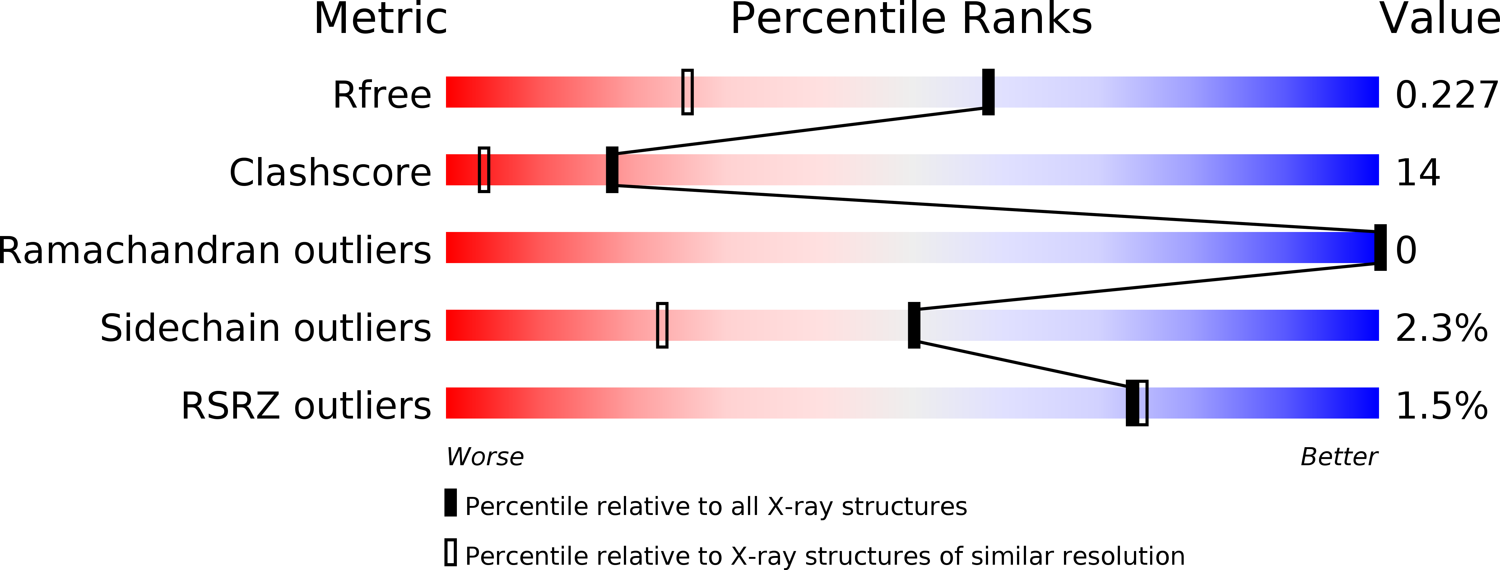
Resolution:
1.65 Å
R-Value Free:
0.22
R-Value Work:
0.17
R-Value Observed:
0.17
Space Group:
P 41 21 2
Macromolecular Entities
Polymer Type:polypeptide(L)
Molecule:Golgi reassembly-stacking protein 2
Gene (Uniprot):GORASP2
Chain IDs:A
Chain Length:209
Number of Molecules:1
Biological Source:Homo sapiens
Modified Residue
Compound ID Chain ID Parent Comp ID Details 2D Image
MSE A MET SELENOMETHIONINE
Primary Citation
Structure of the Membrane-tethering GRASP Domain Reveals a Unique PDZ Ligand Interaction That Mediates Golgi Biogenesis.
J.Biol.Chem. 286 20125 20129 (2011)
PMID: 21515684 DOI: 10.1074/jbc.C111.245324

Abstact

Biogenesis of the ribbon-like membrane network of the mammalian Golgi requires membrane tethering by the conserved GRASP domain in GRASP65 and GRASP55, yet the tethering mechanism is not fully understood. Here, we report the crystal structure of the GRASP55 GRASP domain, which revealed an unusual arrangement of two tandem PDZ folds that more closely resemble prokaryotic PDZ domains. Biochemical and functional data indicated that the interaction between the ligand-binding pocket of PDZ1 and an internal ligand on PDZ2 mediates the GRASP self-interaction, and structural analyses suggest that this occurs via a unique mode of internal PDZ ligand recognition. Our data uncover the structural basis for ligand specificity and provide insight into the mechanism of GRASP-dependent membrane tethering of analogous Golgi cisternae.

Legend

Protein

Chemical

Disease

Primary Citation of related structures