3RLD image
Deposition Date 2011-04-19
Release Date 2012-02-08
Last Version Date 2023-09-13
Entry Detail
PDB ID:
3RLD
Keywords:
Title:
Crystal structure of the Y7I mutant of human carbonic anhydrase II
Biological Source:
Source Organism:
Homo sapiens (Taxon ID: 9606)
Host Organism:
Method Details:
Experimental Method:
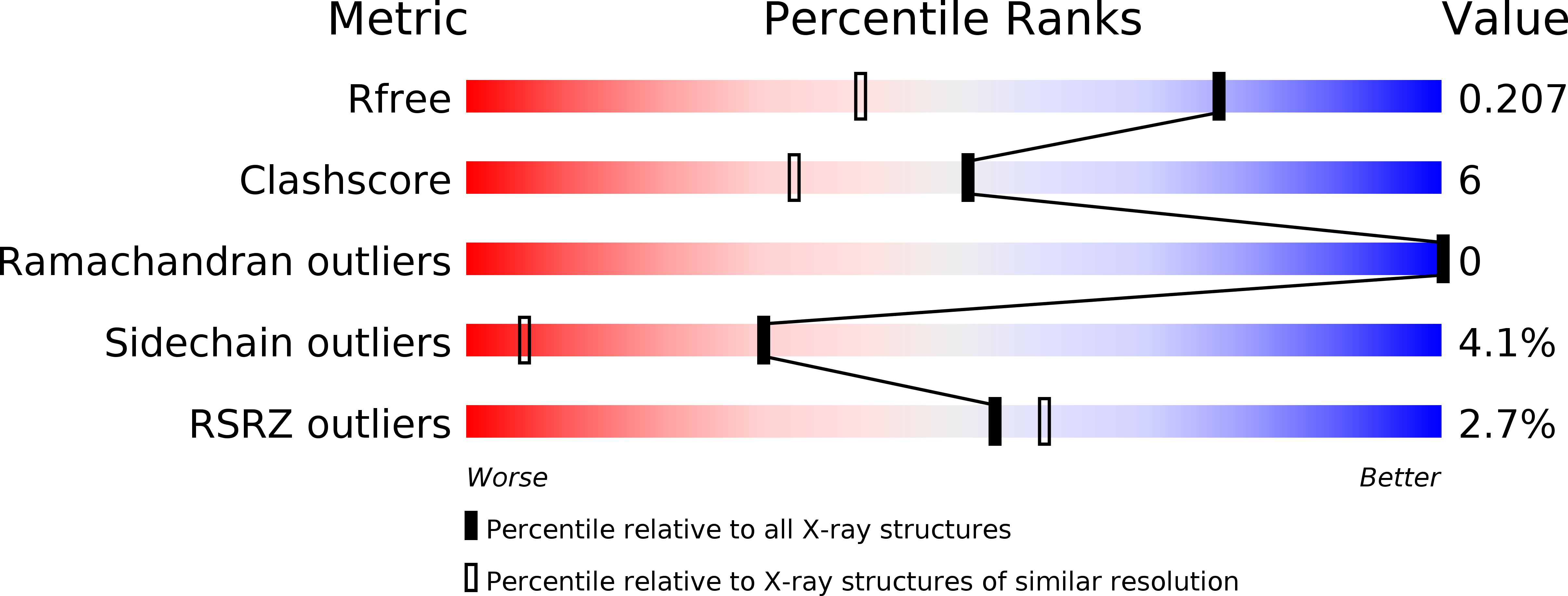
Resolution:
1.50 Å
R-Value Free:
0.21
R-Value Work:
0.16
R-Value Observed:
0.16
Space Group:
P 1 21 1
Macromolecular Entities
Structures with similar UniProt ID
Protein Blast
Polymer Type:polypeptide(L)
Molecule:Carbonic anhydrase 2
Gene (Uniprot):CA2
Mutagens:Y7I
Chain IDs:A
Chain Length:260
Number of Molecules:1
Biological Source:Homo sapiens
Primary Citation
Kinetic and crystallographic studies of the role of tyrosine 7 in the active site of human carbonic anhydrase II.
Arch.Biochem.Biophys. 506 181 187 (2011)
PMID: 21145876 DOI: 10.1016/j.abb.2010.12.004

Abstact

The rate limiting step in catalysis of bicarbonate dehydration by human carbonic anhydrase II (HCA II) is an intramolecular proton transfer from His64 to the zinc-bound hydroxide. We have examined the role of Tyr7 using site-specific mutagenesis and measuring catalysis by the ¹⁸O exchange method using membrane inlet mass spectrometry. The side chain of Tyr7 in HCA II extends into the active-site cavity about 7 Å from the catalytic zinc atom. Replacement of Tyr7 with eight other amino acids had no effect on the interconversion of bicarbonate and CO₂, but in some cases caused enhancements in the rate constant of proton transfer by nearly 10-fold. The variant Y7I HCA II enhanced intramolecular proton transfer approximately twofold; its structure was determined by X-ray crystallography at 1.5 Å resolution. No changes were observed in the ordered solvent structure in the active-site cavity or in the conformation of the side chain of the proton shuttle His64. However, the first 11 residues of the amino-terminal chain in Y7I HCA II assumed an alternate conformation compared with the wild type. Differential scanning calorimetry showed variants at position 7 had a melting temperature approximately 8 °C lower than that of the wild type.

Legend

Protein

Chemical

Disease

Primary Citation of related structures
Feedback Form
Name
Email
Institute
Feedback