
Deposition Date
2011-04-19
Release Date
2011-09-28
Last Version Date
2024-02-28
Entry Detail
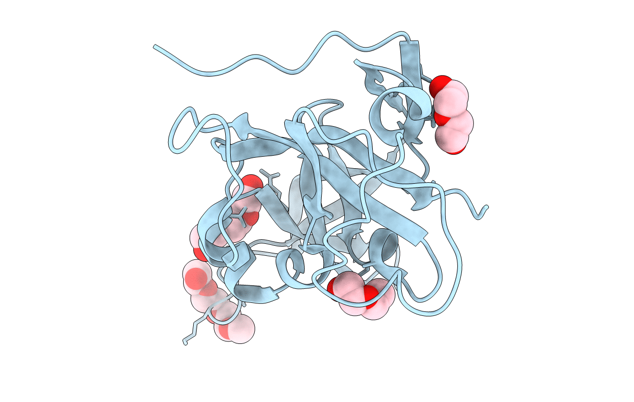
PDB ID:
3RLC
Keywords:
Title:
Crystal structure of the read-through domain from bacteriophage Qbeta A1 protein, hexagonal crystal form
Biological Source:
Source Organism(s):
Enterobacteria phage Qbeta (Taxon ID: 39803)
Expression System(s):
Method Details:
Experimental Method:
Resolution:
2.90 Å
R-Value Free:
0.29
R-Value Work:
0.21
R-Value Observed:
0.21
Space Group:
P 63 2 2
