3RK0 image
Deposition Date 2011-04-16
Release Date 2011-05-11
Last Version Date 2024-10-30
Entry Detail
PDB ID:
3RK0
Keywords:
Title:
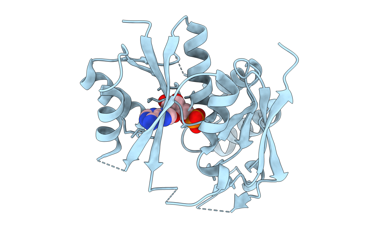
X-ray crystal Structure of the putative N-type ATP pyrophosphatase (PF0828) in complex with AMP from Pyrococcus furiosus, Northeast Structural Genomics Consortium Target PfR23
Biological Source:
Source Organism(s):
Expression System(s):
Method Details:
Experimental Method:
Resolution:
2.40 Å
R-Value Free:
0.27
R-Value Work:
0.22
R-Value Observed:
0.22
Space Group:
P 41 21 2
Macromolecular Entities
Structures with similar UniProt ID
Protein Blast
Polymer Type:polypeptide(L)
Molecule:N-type ATP pyrophosphatase superfamily
Gene (Uniprot):PF0828
Chain IDs:A
Chain Length:237
Number of Molecules:1
Biological Source:Pyrococcus furiosus
Modified Residue
Compound ID Chain ID Parent Comp ID Details 2D Image
MSE A MET SELENOMETHIONINE
Ligand Molecules
Primary Citation
A large conformational change in the putative ATP pyrophosphatase PF0828 induced by ATP binding.
Acta Crystallogr.,Sect.F 67 1323 1327 (2011)
PMID: 22102225 DOI: 10.1107/S1744309111031447

Abstact

ATP pyrophosphatases (ATP PPases) are widely distributed in archaea and eukaryotes. They share an HUP domain at the N-terminus with a conserved PP-motif that interacts with the phosphates of ATP. The PF0828 protein from Pyrococcus furiosus is a member of the ATP PPase superfamily and it also has a 100-residue C-terminal extension that contains a strictly conserved EGG(E/D)xE(T/S) motif, which has been named the EGT-motif. Here, crystal structures of PF0828 alone and in complex with ATP or AMP are reported. The HUP domain contains a central five-stranded β-sheet that is surrounded by four helices, as in other related structures. The C-terminal extension forms a separate domain, named the EGT domain, which makes tight interactions with the HUP domain, bringing the EGT-motif near to the PP-motif and defining the putative active site of PF0828. Both motifs interact with the phosphate groups of ATP. A loop in the HUP domain undergoes a large conformational change to recognize the adenine base of ATP. In solution and in the crystal PF0828 is a dimer formed by the side-by-side arrangement of the HUP domains of the two monomers. The putative active site is located far from the dimer interface.

Legend

Protein

Chemical

Disease

Primary Citation of related structures
Feedback Form
Name
Email
Institute
Feedback