3RHX image
Deposition Date 2011-04-12
Release Date 2011-05-04
Last Version Date 2024-02-28
Entry Detail
PDB ID:
3RHX
Title:
Crystal structure of the catalytic domain of FGFR1 kinase in complex with ARQ 069
Biological Source:
Source Organism:
Homo sapiens (Taxon ID: 9606)
Host Organism:
Method Details:
Experimental Method:
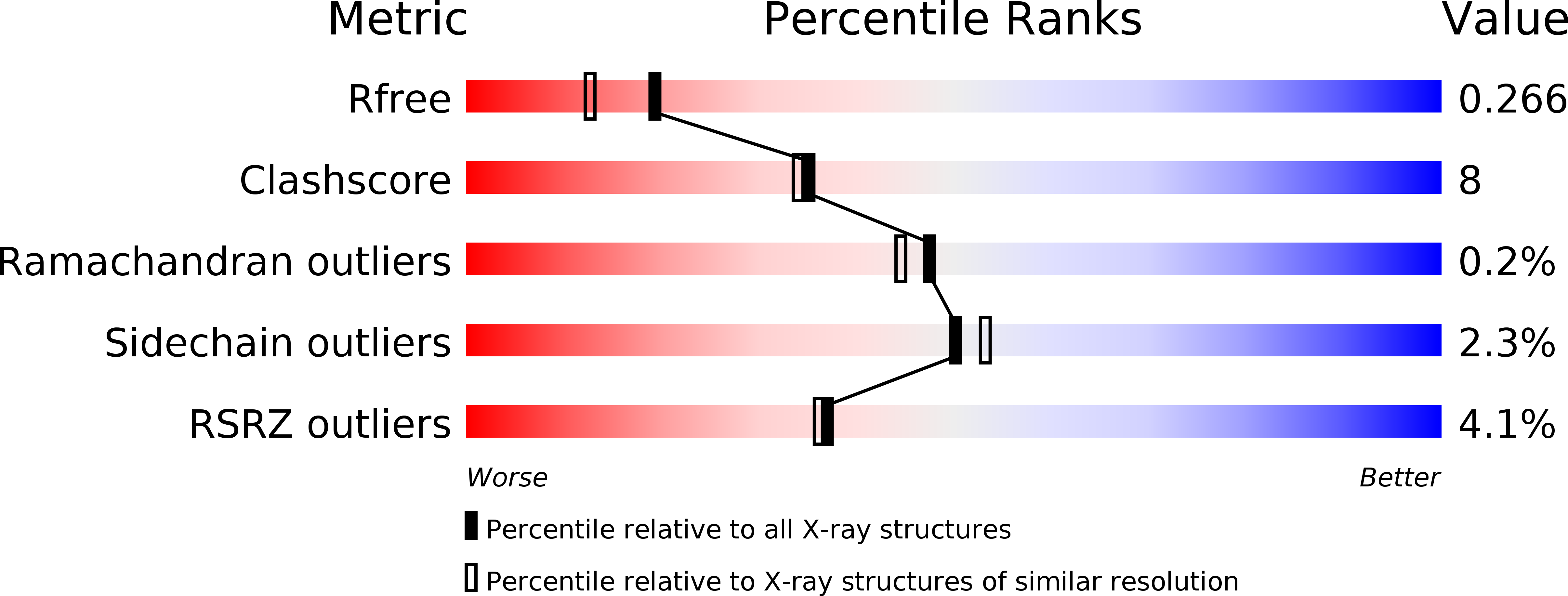
Resolution:
2.01 Å
R-Value Free:
0.26
R-Value Work:
0.20
R-Value Observed:
0.20
Space Group:
C 1 2 1
Macromolecular Entities
Structures with similar UniProt ID
Protein Blast
Polymer Type:polypeptide(L)
Molecule:Basic fibroblast growth factor receptor 1
Gene (Uniprot):FGFR1
Mutagens:C488A, C584S
Chain IDs:A (auth: B), B (auth: A)
Chain Length:306
Number of Molecules:2
Biological Source:Homo sapiens
Primary Citation
A novel mode of protein kinase inhibition exploiting hydrophobic motifs of autoinhibited kinases: discovery of ATP-independent inhibitors of fibroblast growth factor receptor.
J.Biol.Chem. 286 20677 20687 (2011)
PMID: 21454610 DOI: 10.1074/jbc.M110.213736

Abstact

Protein kinase inhibitors with enhanced selectivity can be designed by optimizing binding interactions with less conserved inactive conformations because such inhibitors will be less likely to compete with ATP for binding and therefore may be less impacted by high intracellular concentrations of ATP. Analysis of the ATP-binding cleft in a number of inactive protein kinases, particularly in the autoinhibited conformation, led to the identification of a previously undisclosed non-polar region in this cleft. This ATP-incompatible hydrophobic region is distinct from the previously characterized hydrophobic allosteric back pocket, as well as the main pocket. Generalized hypothetical models of inactive kinases were constructed and, for the work described here, we selected the fibroblast growth factor receptor (FGFR) tyrosine kinase family as a case study. Initial optimization of a FGFR2 inhibitor identified from a library of commercial compounds was guided using structural information from the model. We describe the inhibitory characteristics of this compound in biophysical, biochemical, and cell-based assays, and have characterized the binding mode using x-ray crystallographic studies. The results demonstrate, as expected, that these inhibitors prevent activation of the autoinhibited conformation, retain full inhibitory potency in the presence of physiological concentrations of ATP, and have favorable inhibitory activity in cancer cells. Given the widespread regulation of kinases by autoinhibitory mechanisms, the approach described herein provides a new paradigm for the discovery of inhibitors by targeting inactive conformations of protein kinases.

Legend

Protein

Chemical

Disease

Primary Citation of related structures