3RGS image
Entry Detail
PDB ID:
3RGS
Keywords:
Title:
Structural and kinetic analysis of the beef liver catalase with the ammonia as a ligand
Biological Source:
Source Organism:
PDB Version:
Deposition Date:
2011-04-08
Release Date:
2011-05-11
Method Details:
Experimental Method:
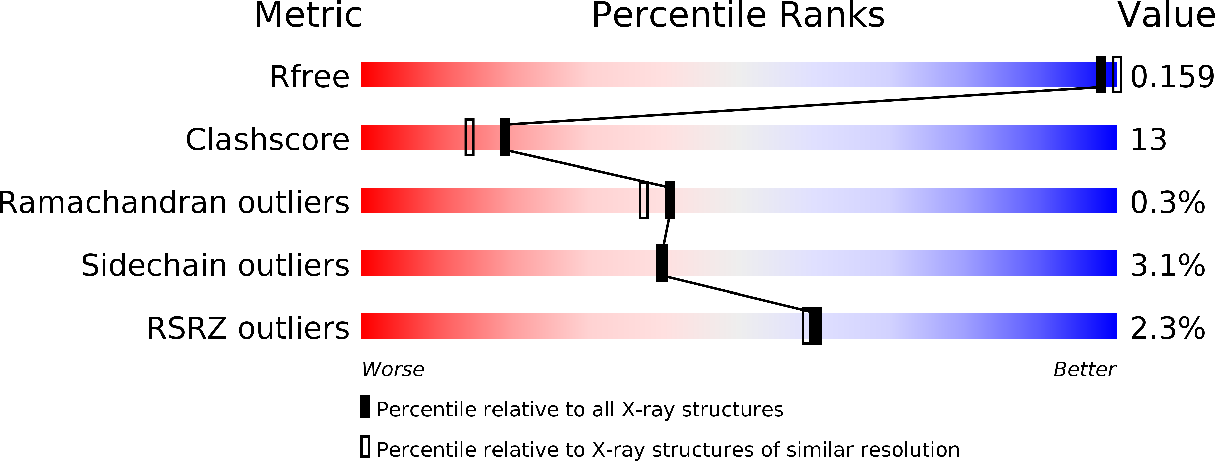
Resolution:
1.99 Å
R-Value Free:
0.19
R-Value Work:
0.16
R-Value Observed:
0.16
Space Group:
P 21 21 21
Macromolecular Entities
Polymer Type:polypeptide(L)
Description:Catalase
Chain IDs:A, B, C, D
Chain Length:499
Number of Molecules:4
Biological Source:Bos taurus
Primary Citation
Interaction of nitric oxide with catalase: structural and kinetic analysis.
Biochemistry 50 4491 4503 (2011)
PMID: 21524057 DOI: 10.1021/bi200130r

Abstact

We present the structures of bovine catalase in its native form and complexed with ammonia and nitric oxide, obtained by X-ray crystallography. Using the NO generator 1-(N,N-diethylamino)diazen-1-ium-1,2-diolate, we were able to generate sufficiently high NO concentrations within the catalase crystals that substantial occupation was observed despite a high dissociation rate. Nitric oxide seems to be slightly bent from the heme normal that may indicate some iron(II) character in the formally ferric catalase. Microspectrophotometric investigations inline with the synchrotron X-ray beam reveal photoreduction of the central heme iron. In the cases of the native and ammonia-complexed catalase, reduction is accompanied by a relaxation phase. This is likely not the case for the catalase NO complex. The kinetics of binding of NO to catalase were investigated using NO photolyzed from N,N'-bis(carboxymethyl)-N,N'-dinitroso-p-phenylenediamine using an assay that combines catalase with myoglobin binding kinetics. The off rate is 1.5 s(-1). Implications for catalase function are discussed.

Legend

Protein

Chemical

Disease

Primary Citation of related structures