3RDO image
Deposition Date 2011-04-01
Release Date 2011-07-06
Last Version Date 2023-09-13
Entry Detail
PDB ID:
3RDO
Title:
Crystal structure of R7-2 streptavidin complexed with biotin
Biological Source:
Source Organism:
Host Organism:
Method Details:
Experimental Method:
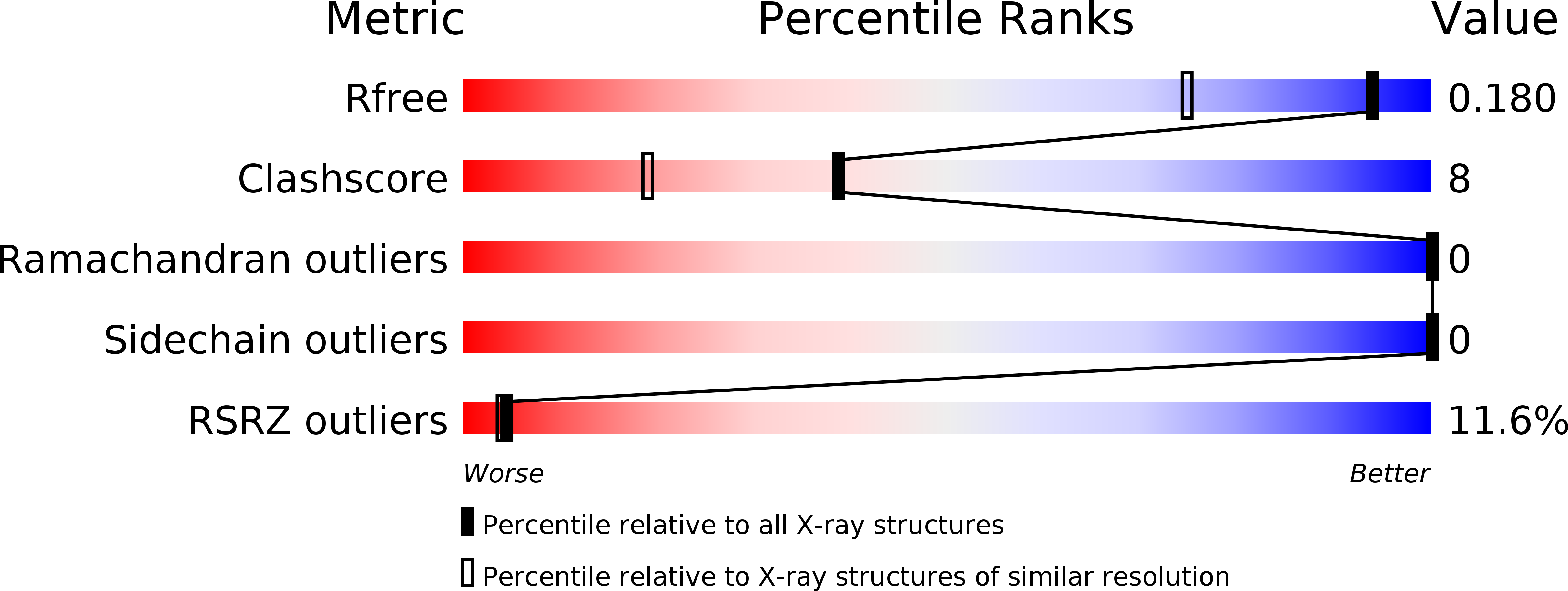
Resolution:
1.40 Å
R-Value Free:
0.17
R-Value Work:
0.15
R-Value Observed:
0.15
Space Group:
I 41 2 2
Macromolecular Entities
Polymer Type:polypeptide(L)
Molecule:Streptavidin
Mutations:T90S, W108V, L110T, F29L, S52G, R53S
Chain IDs:A
Chain Length:153
Number of Molecules:1
Biological Source:Streptomyces avidinii
Primary Citation
Evolved streptavidin mutants reveal key role of loop residue in high-affinity binding.
Protein Sci. 20 1145 1154 (2011)
PMID: 21520321 DOI: 10.1002/pro.642

Abstact

We have performed a detailed analysis of streptavidin variants with altered specificity towards desthiobiotin. In addition to changes in key residues which widen the ligand binding pocket and accommodate the more structurally flexible desthiobiotin, the data revealed the role of a key, non-active site mutation at the base of the flexible loop (S52G) which slows dissociation of this ligand by approximately sevenfold. Our data suggest that this mutation results in the loss of a stabilizing contact which keeps this loop open and accessible in the absence of ligand. When this mutation was introduced into the wild-type protein, destabilization of the opened loop conferred a ∼10-fold decrease in both the on-rate and off-rate for the ligand biotin-4-fluoroscein. A similar effect was observed when this mutation was added to a monomeric form of this protein. Our results provide key insight into the role of the streptavidin flexible loop in ligand binding and maintaining high affinity interactions.

Legend

Protein

Chemical

Disease

Primary Citation of related structures