
Deposition Date
1998-02-24
Release Date
1999-02-23
Last Version Date
2024-05-22
Entry Detail
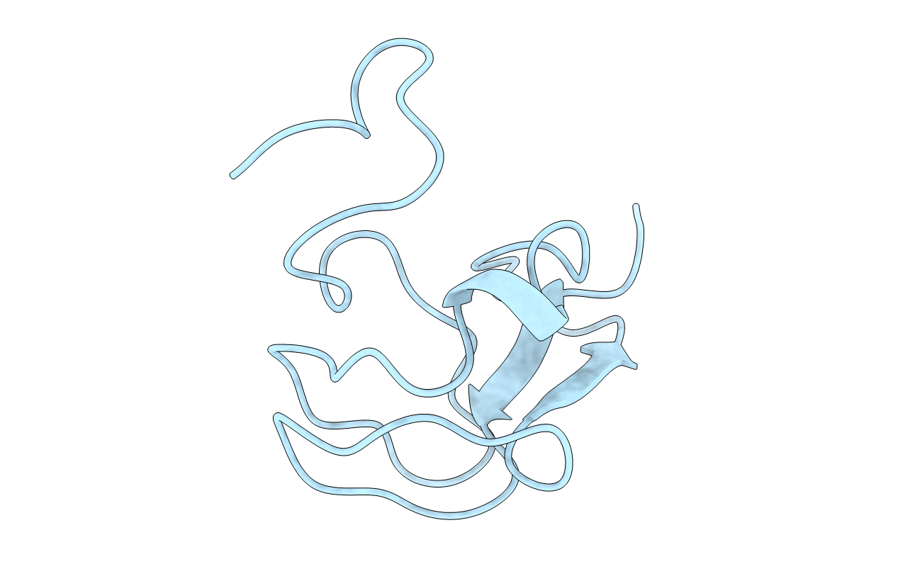
PDB ID:
3RDN
Keywords:
Title:
NMR STRUCTURE OF THE N-TERMINAL DOMAIN WITH A LINKER PORTION OF ANTARCTIC EEL POUT ANTIFREEZE PROTEIN RD3, MINIMIZED AVERAGE STRUCTURE
Biological Source:
Source Organism:
Lycodichthys dearborni (Taxon ID: 8201)
Host Organism:
Method Details:
Experimental Method:
Conformers Calculated:
40
Conformers Submitted:
1
Selection Criteria:
AVERAGE
