Deposition Date
2011-03-30
Release Date
2012-03-21
Last Version Date
2024-03-20
Entry Detail
PDB ID:
3RBY
Keywords:
Title:
Crystal structure of uncharacterized protein YLR301w from Saccharomycces cerevisiae
Biological Source:
Source Organism(s):
Saccharomyces cerevisiae (Taxon ID: 4932)
Expression System(s):
Method Details:
Experimental Method:
Resolution:
2.30 Å
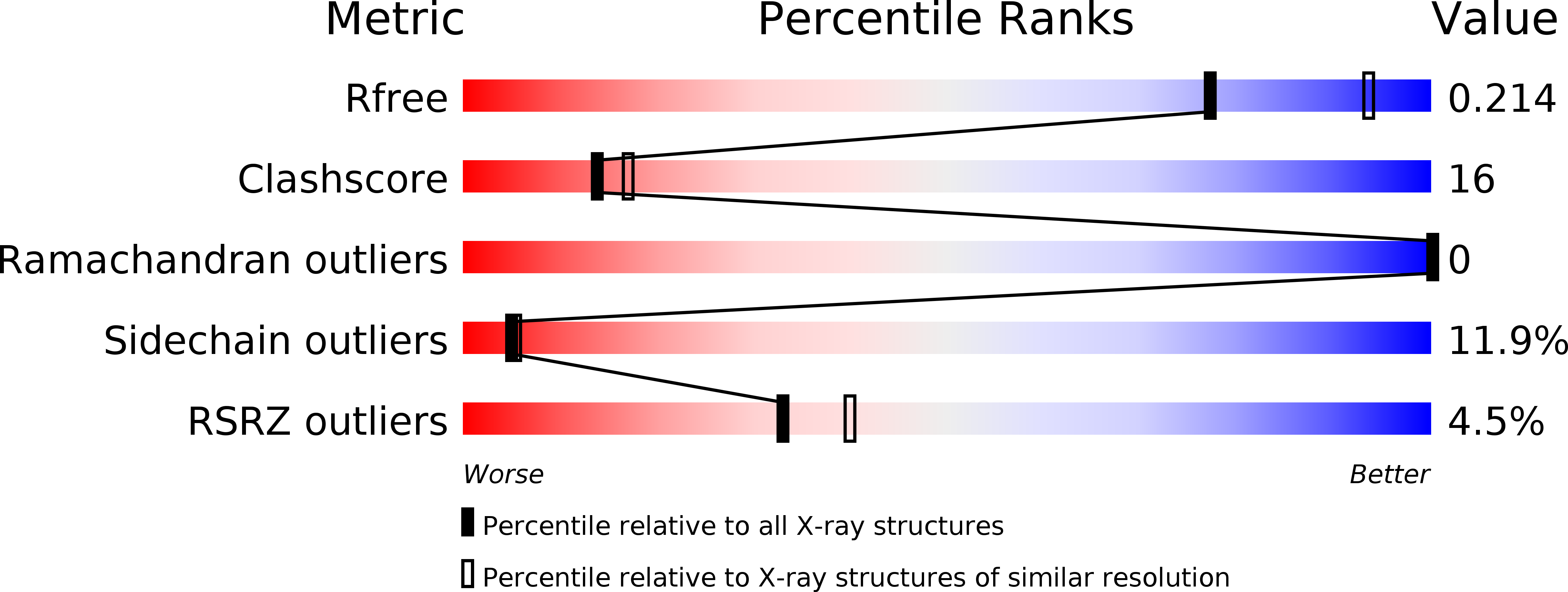
R-Value Free:
0.24
R-Value Work:
0.20
R-Value Observed:
0.20
Space Group:
P 65 2 2