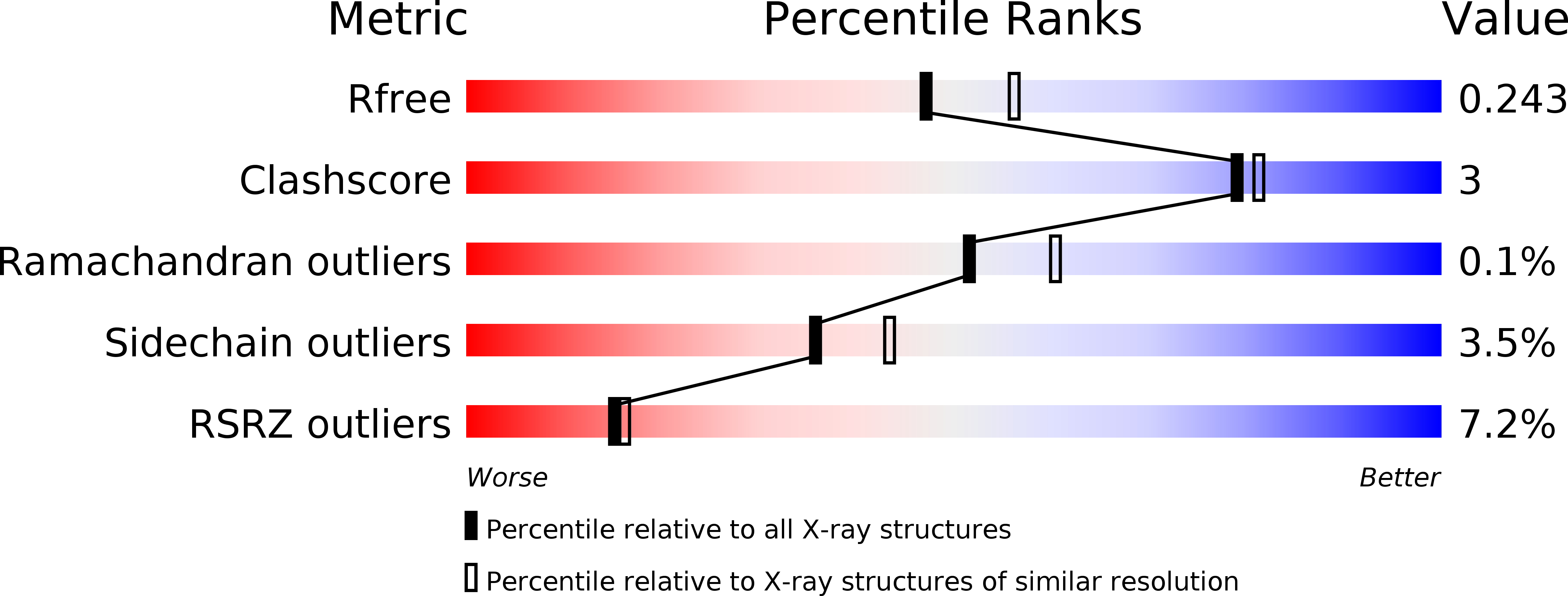
3RAQ image
Deposition Date 2011-03-28
Release Date 2011-06-15
Last Version Date 2023-09-13
Entry Detail
PDB ID:
3RAQ
Keywords:
Title:
Dpo4 extension ternary complex with 3'-terminal primer C base opposite the 1-methylguanine (MG1) lesion
Biological Source:
Source Organism:
Sulfolobus solfataricus (Taxon ID: 2287)
(Taxon ID: )
Host Organism:
Method Details:
Experimental Method:
Resolution:
2.25 Å
R-Value Free:
0.23
R-Value Work:
0.19
R-Value Observed:
0.19
Space Group:
P 1
Macromolecular Entities
Polymer Type:polypeptide(L)
Molecule:DNA polymerase IV
Gene (Uniprot):dbh
Chain IDs:A, D (auth: B)
Chain Length:341
Number of Molecules:2
Biological Source:Sulfolobus solfataricus
Polymer Type:polydeoxyribonucleotide
Molecule:DNA (5'-D(*GP*TP*TP*GP*GP*AP*TP*GP*GP*TP*AP*GP*(DOC))-3')
Chain IDs:B (auth: D), E (auth: H)
Chain Length:13
Number of Molecules:2
Biological Source:
Polymer Type:polydeoxyribonucleotide
Molecule:DNA (5'-D(*C*CP*TP*AP*AP*CP*(MG1)P*CP*TP*AP*CP*CP*AP*TP*CP*CP*AP*AP*CP*C)-3')
Chain IDs:C (auth: E), F (auth: J)
Chain Length:20
Number of Molecules:2
Biological Source:
Modified Residue
Compound ID Chain ID Parent Comp ID Details 2D Image
DOC B DC 2',3'-DIDEOXYCYTIDINE-5'-MONOPHOSPHATE
MG1 C DG ?
Primary Citation
Implications for damage recognition during Dpo4-mediated mutagenic bypass of m1G and m3C lesions.
Structure 19 821 832 (2011)
PMID: 21645853 DOI: 10.1016/j.str.2011.03.020

Abstact

DNA is susceptible to alkylation damage by a number of environmental agents that modify the Watson-Crick edge of the bases. Such lesions, if not repaired, may be bypassed by Y-family DNA polymerases. The bypass polymerase Dpo4 is strongly inhibited by 1-methylguanine (m1G) and 3-methylcytosine (m3C), with nucleotide incorporation opposite these lesions being predominantly mutagenic. Further, extension after insertion of both correct and incorrect bases, introduces additional base substitution and deletion errors. Crystal structures of the Dpo4 ternary extension complexes with correct and mismatched 3'-terminal primer bases opposite the lesions reveal that both m1G and m3C remain positioned within the DNA template/primer helix. However, both correct and incorrect pairing partners exhibit pronounced primer terminal nucleotide distortion, being primarily evicted from the DNA helix when opposite m1G or misaligned when pairing with m3C. Our studies provide insights into mechanisms related to hindered and mutagenic bypass of methylated lesions and models associated with damage recognition by repair demethylases.

Legend

Protein

Chemical

Disease

Primary Citation of related structures