Deposition Date
2011-03-25
Release Date
2011-08-17
Last Version Date
2023-09-13
Entry Detail
PDB ID:
3R9I
Keywords:
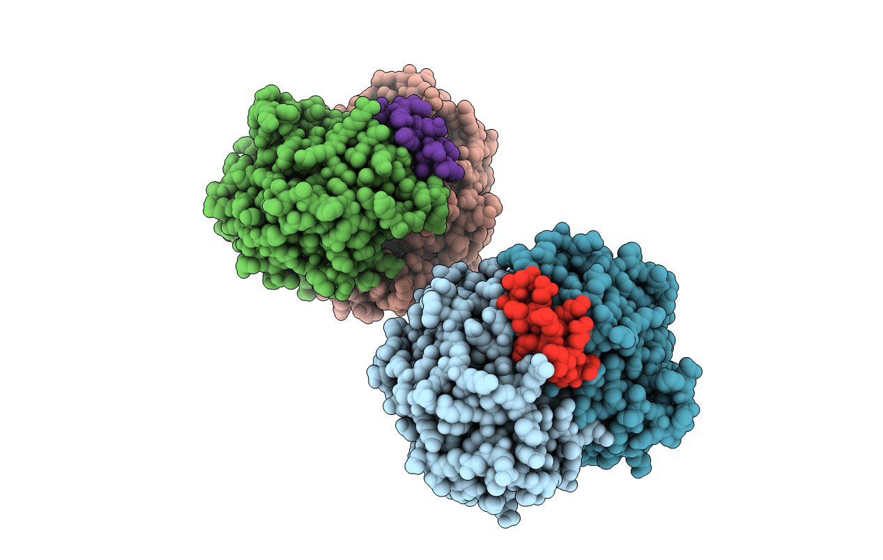
Title:
2.6A resolution structure of MinD complexed with MinE (12-31) peptide
Biological Source:
Source Organism(s):
Escherichia coli (Taxon ID: 83333)
Expression System(s):
Method Details:
Experimental Method:
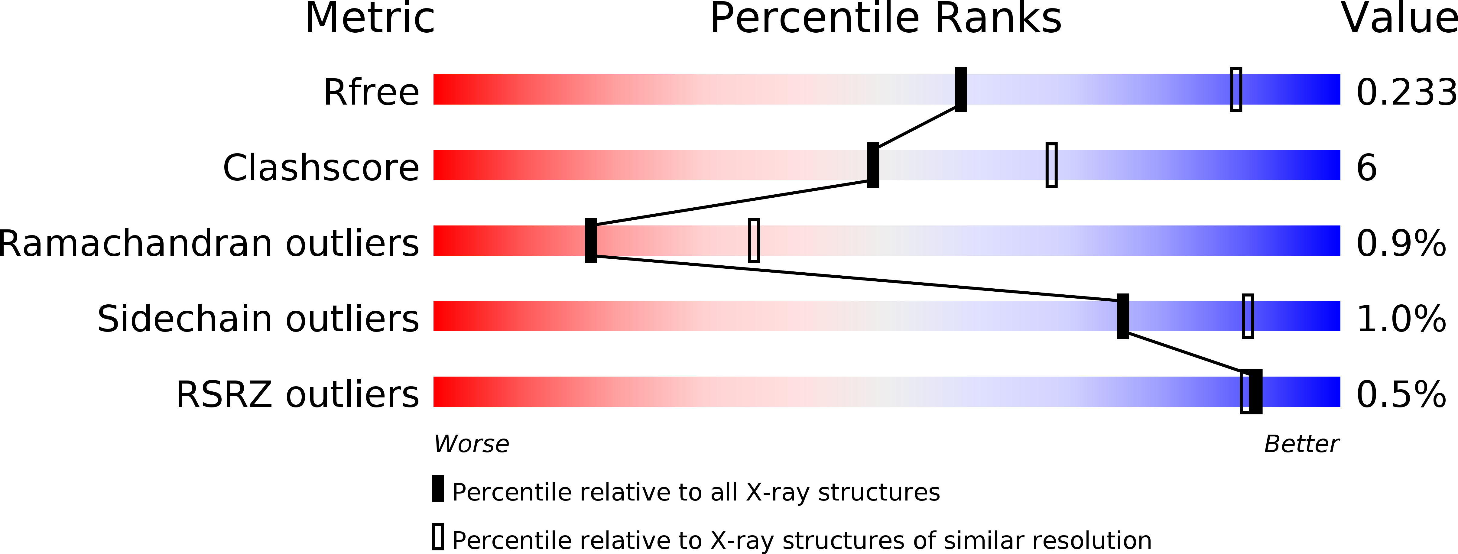
Resolution:
2.60 Å
R-Value Free:
0.24
R-Value Work:
0.20
R-Value Observed:
0.20
Space Group:
P 1