Deposition Date
2011-03-18
Release Date
2012-01-11
Last Version Date
2023-09-13
Entry Detail
PDB ID:
3R5G
Keywords:
Title:
Crystal structure of the adenylyl cyclase CyaB from P. aeruginosa
Biological Source:
Source Organism(s):
Pseudomonas aeruginosa (Taxon ID: 287)
Expression System(s):
Method Details:
Experimental Method:
Resolution:
1.50 Å
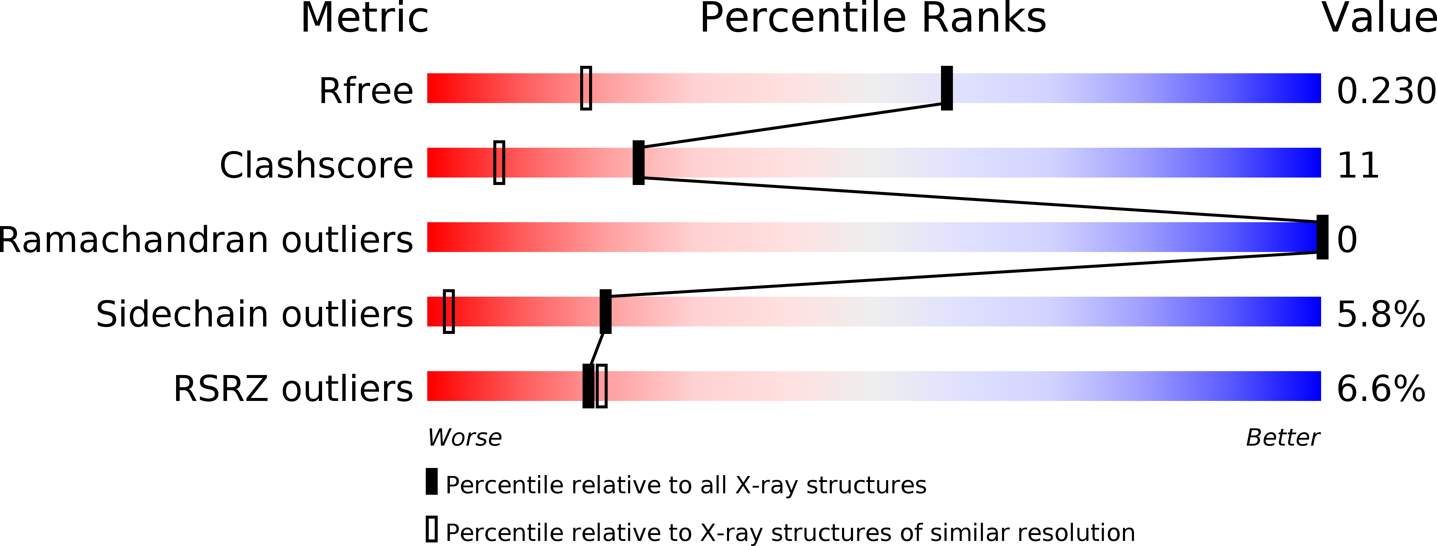
R-Value Free:
0.23
R-Value Work:
0.18
R-Value Observed:
0.18
Space Group:
P 1 2 1