
Deposition Date
2011-03-18
Release Date
2012-02-01
Last Version Date
2024-03-20
Entry Detail
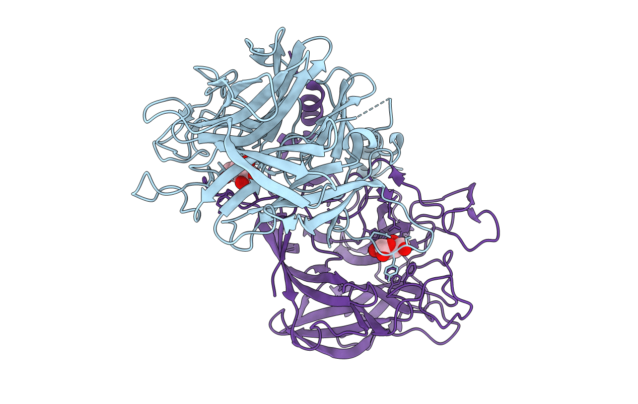
PDB ID:
3R4Z
Keywords:
Title:
Crystal structure of alpha-neoagarobiose hydrolase (ALPHA-NABH) in complex with alpha-d-galactopyranose from Saccharophagus degradans 2-40
Biological Source:
Source Organism(s):
Saccharophagus degradans (Taxon ID: 203122)
Expression System(s):
Method Details:
Experimental Method:
Resolution:
1.55 Å
R-Value Free:
0.18
R-Value Work:
0.16
R-Value Observed:
0.16
Space Group:
C 1 2 1
