3R4H image
Deposition Date 2011-03-17
Release Date 2011-11-16
Last Version Date 2025-03-26
Entry Detail
PDB ID:
3R4H
Keywords:
Title:
Crystal structure of the 4-helix coiled coil CC-Tet-phi22
Biological Source:
Source Organism:
Method Details:
Experimental Method:
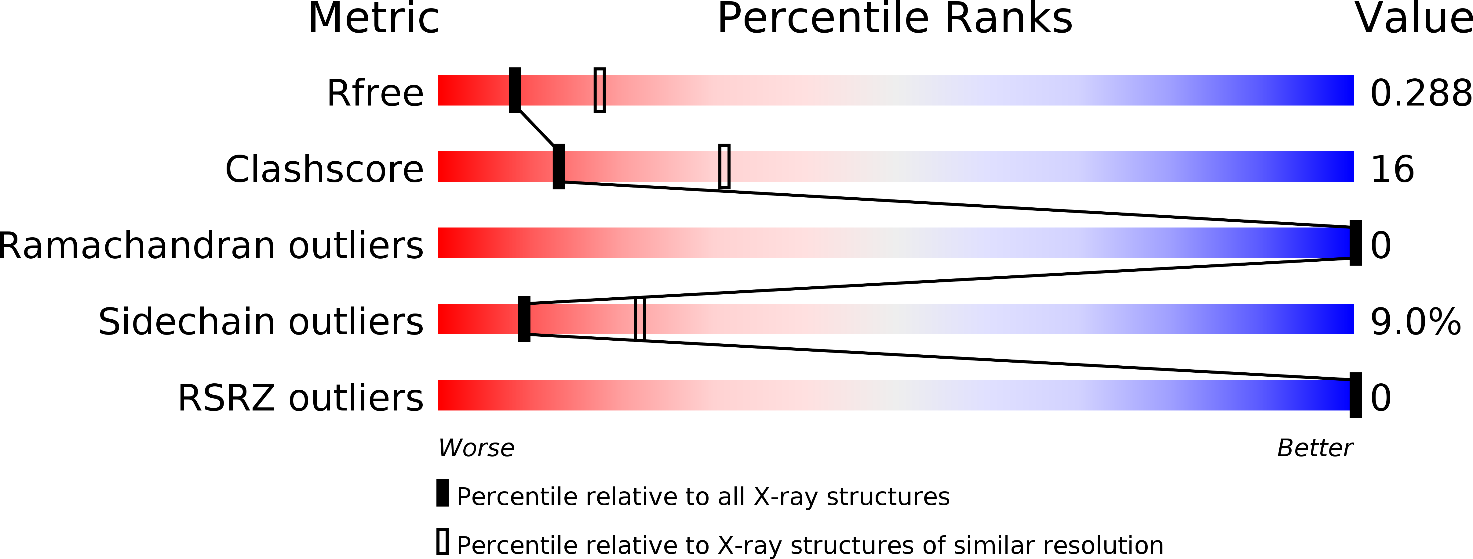
Resolution:
2.70 Å
R-Value Free:
0.27
R-Value Work:
0.20
R-Value Observed:
0.20
Space Group:
P 43 21 2
Macromolecular Entities
Polymer Type:polypeptide(L)
Molecule:coiled coil helix CC-Tet-phi22
Chain IDs:A, B, C, D, E, F
Chain Length:34
Number of Molecules:6
Biological Source:Synthetic construct
Modified Residue
Compound ID Chain ID Parent Comp ID Details 2D Image
PHI A PHE IODO-PHENYLALANINE
Primary Citation
A de novo peptide hexamer with a mutable channel.
Nat.Chem.Biol. 7 935 941 (2011)
PMID: 22037471 DOI: 10.1038/nchembio.692

Abstact

The design of new proteins that expand the repertoire of natural protein structures represents a formidable challenge. Success in this area would increase understanding of protein structure and present new scaffolds that could be exploited in biotechnology and synthetic biology. Here we describe the design, characterization and X-ray crystal structure of a new coiled-coil protein. The de novo sequence forms a stand-alone, parallel, six-helix bundle with a channel running through it. Although lined exclusively by hydrophobic leucine and isoleucine side chains, the 6-Å channel is permeable to water. One layer of leucine residues within the channel is mutable, accepting polar aspartic acid and histidine side chains, which leads to subdivision and organization of solvent within the lumen. Moreover, these mutants can be combined to form a stable and unique (Asp-His)(3) heterohexamer. These new structures provide a basis for engineering de novo proteins with new functions.

Legend

Protein

Chemical

Disease

Primary Citation of related structures