Deposition Date
2011-03-17
Release Date
2011-06-22
Last Version Date
2024-10-16
Entry Detail
PDB ID:
3R4D
Keywords:
Title:
Crystal structure of mouse coronavirus receptor-binding domain complexed with its murine receptor
Biological Source:
Source Organism(s):
Mus musculus (Taxon ID: 10090)
Murine coronavirus (Taxon ID: 694005)
Murine coronavirus (Taxon ID: 694005)
Expression System(s):
Method Details:
Experimental Method:
Resolution:
3.10 Å
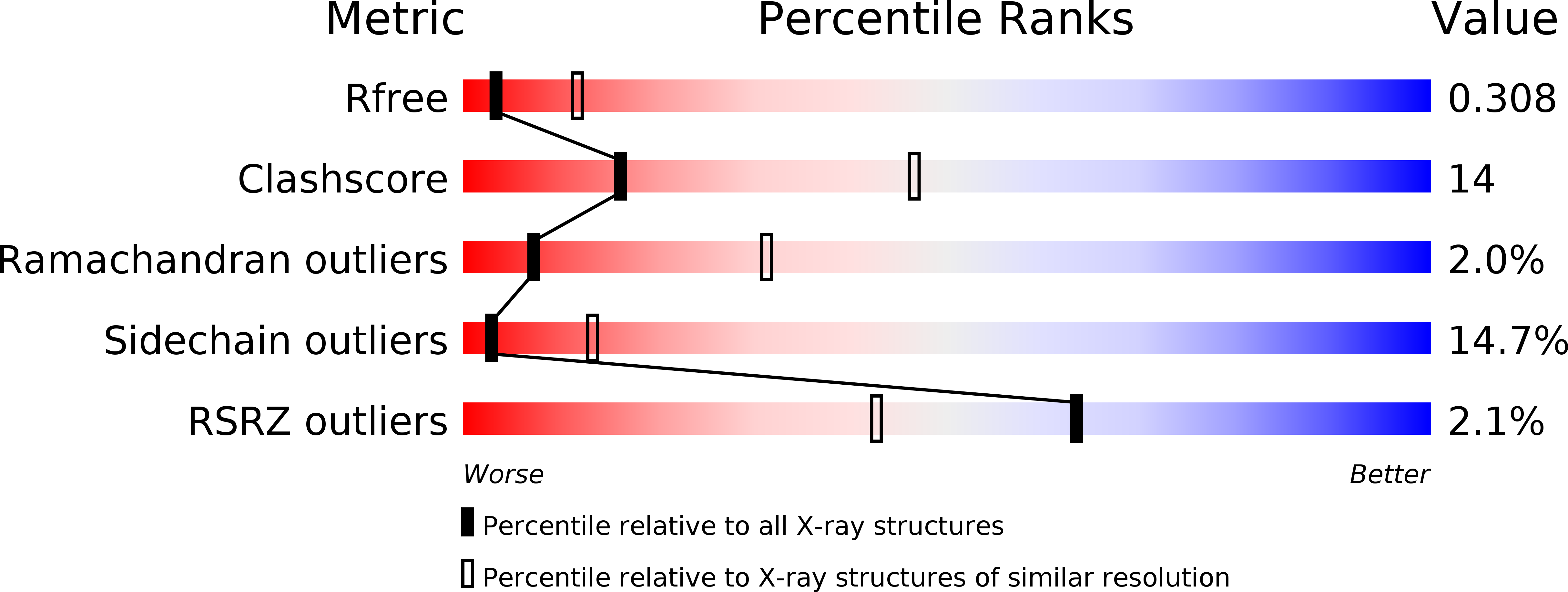
R-Value Free:
0.30
R-Value Work:
0.24
Space Group:
P 61 2 2